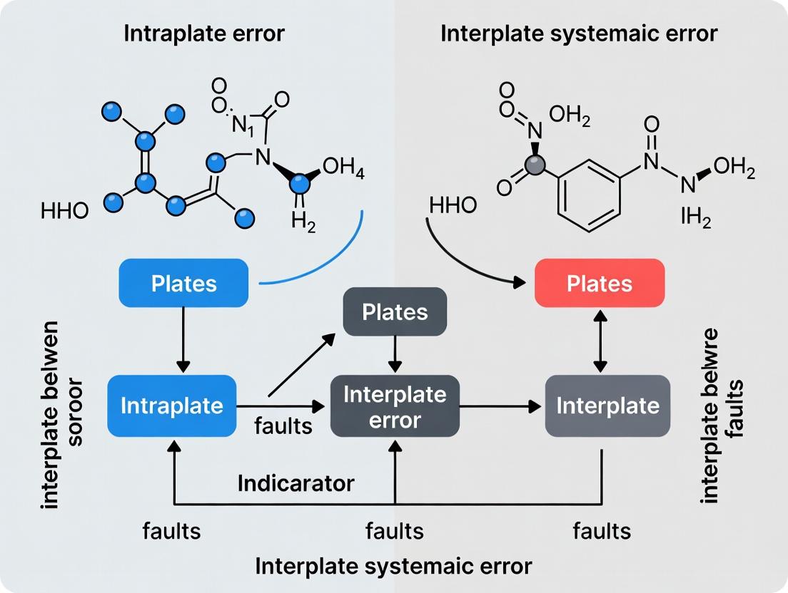
Beyond the Plate: Mastering Intraplate and Interplate Systematic Error in High-Throughput Screening
This article provides a comprehensive guide for researchers and drug development professionals on the critical challenge of systematic error in high-throughput screening (HTS).
Beyond the Plate: Mastering Intraplate and Interplate Systematic Error in High-Throughput Screening
Abstract
This article provides a comprehensive guide for researchers and drug development professionals on the critical challenge of systematic error in high-throughput screening (HTS). It explores the fundamental distinctions between intraplate (spatial variation within a single microtiter plate) and interplate (variation between different plates) systematic errors, detailing their common causes such as robotic handling, environmental gradients, and assay timing[citation:1]. The scope covers methodological approaches for identification and correction, including advanced median filter techniques and computational quality control frameworks[citation:1][citation:7]. It further delves into troubleshooting and optimization strategies for assay design and data processing, and concludes with validation methods using robust statistical metrics like the Z'-factor to assess data quality improvements[citation:2]. The full discussion synthesizes how mastering these errors is essential for improving dynamic range, hit confirmation rates, and the overall reliability of biomedical research data[citation:1].
Decoding the Signal from the Noise: Defining Intraplate vs. Interplate Systematic Error
This technical whitepates a critical framework for systematic error research in high-throughput screening (HTS) and assay development, distinguishing between intraplate (spatial) and interplate (temporal/batch) variation. This core distinction is fundamental for robust assay validation, data normalization, and the reliable identification of bioactive compounds in drug discovery. Within the broader thesis on understanding systematic errors, precise delineation of these variation sources enables targeted mitigation strategies, directly impacting the reproducibility and quality of scientific research.
Core Definitions and Thesis Context
Intraplate Variation refers to systematic spatial biases within a single microtiter plate. These are non-random patterns of measurement error correlated with well position, arising from factors such as edge evaporation effects, temperature gradients across the plate during incubation, pipetting head inaccuracies, or reader optics. It is inherently spatial.
Interplate Variation refers to systematic differences between plates processed at different times or in different batches. This temporal/batch variation stems from reagent lot changes, ambient temperature/humidity shifts, recalibration of instruments, or day-to-day operator differences.
The broader thesis posits that disentangling these two orthogonal dimensions of systematic error is a prerequisite for developing universally applicable normalization and quality control protocols. Effective control of intraplate variation ensures plate homogeneity, while managing interplate variation ensures experimental reproducibility across runs and laboratories.
Quantitative Characterization of Variation
Empirical studies quantify these variations using control compounds (e.g., DMSO blanks, positive/negative controls) replicated across plates and positions. Key metrics include Z'-factor for assay quality, and coefficient of variation (CV) for precision.
Table 1: Typical Quantitative Metrics for Intraplate vs. Interplate Variation
| Metric | Intraplate Variation (Spatial) | Interplate Variation (Temporal/Batch) | Optimal Target |
|---|---|---|---|
| Z'-factor | Calculated per plate using intraplate controls. | Calculated across plates using mean of plate controls. | > 0.5 (Excellent assay) |
| CV of Controls | CV across replicate control wells within a plate. | CV of the plate mean control values across plates/batches. | < 10-20% (Assay-dependent) |
| Signal-to-Noise (S/N) | Ratio for controls within a single plate. | Ratio of plate mean signals across batches. | > 10 (Robust assay) |
| Primary Source | Edge effects, thermal gradients, pipetting drift. | Reagent lot changes, instrument recalibration, environmental drift. | N/A |
Table 2: Experimental Design for Disentangling Variation Sources
| Component | Purpose | Layout Example (96-well) |
|---|---|---|
| Negative Controls | Measures baseline signal and error. | Columns 1 & 12, all rows (n=16). |
| Positive Controls | Measures maximal signal and error. | Columns 2 & 11, all rows (n=16). |
| Spatial Control Grid | Maps intraplate gradients. | DMSO in all wells of plates designated for variation mapping. |
| Interplate Reference | Anchors batch normalization. | At least one standardized control plate per batch run. |
Experimental Protocols for Assessment
Protocol 3.1: Comprehensive Intraplate Variation Mapping
Objective: To visualize and quantify spatial bias on a specific instrument-platform-reagent set.
- Plate Preparation: Prepare a minimum of three replicate plates. Fill all wells with an identical solution containing a reporter (e.g., fluorophore at mid-range intensity) in assay buffer.
- Instrument Reading: Read plates using the standard assay protocol (e.g., fluorescence, luminescence).
- Data Analysis: For each plate, calculate the mean and standard deviation for the entire plate. Create a heat map of raw signal values. Perform ANOVA with well row and column as factors to quantify significance of positional effects.
- Normalization Test: Apply candidate spatial normalization algorithms (e.g., B-score, loess smoothing, median polish) and re-evaluate heat maps.
Protocol 3.2: Systematic Interplate (Batch) Variation Assessment
Objective: To quantify run-to-run variability over an extended period.
- Longitudinal Study Design: Include an identical set of controls (negative, positive, mid-point reference) on every plate in every batch. Use a standardized plate layout.
- Batch Execution: Run assay plates over multiple days, weeks, or using different reagent lots. Maintain consistent protocol documentation.
- Data Analysis: For each control type, plot the plate mean value over time (run sequence). Calculate the inter-plate CV for each control type. Use control charts (e.g., Levey-Jennings) to identify out-of-control batches.
- Normalization: Apply batch correction methods (e.g., plate mean centering, robust Z-score normalization using batch controls) and reassess interplate CV.
Signaling Pathways & Systematic Error Relationships
Diagram 1: Taxonomy of Systematic Error in HTS (77 chars)
Diagram 2: Systematic Error Mitigation Workflow (81 chars)
The Scientist's Toolkit: Essential Research Reagents & Materials
Table 3: Key Reagent Solutions for Variation Research
| Item | Function | Application in Variation Studies |
|---|---|---|
| DMSO (High-Purity, Low-Hygroscopic) | Universal solvent for compound libraries. | Serves as the standard negative control. Batch consistency is critical for interplate studies. |
| Validated Control Agonist/Antagonist | Pharmacologically active reference compound. | Serves as positive control for calculating Z'-factor and monitoring interplate assay performance. |
| Fluorescent/Luminescent Tracer Plate | Plate with homogeneous signal generation. | A dedicated plate (e.g., with fluorophore in buffer) for mapping intraplate reader and optics bias. |
| Cell Line with Stable Reporter (e.g., Luciferase) | Genetically engineered cellular reagent. | Provides a consistent biological signal source for longitudinal interplate variation studies. |
| Assay-Ready Cryopreserved Cells | Standardized, batch-produced cells. | Minimizes biological variability as a source of interplate variation, isolating technical error. |
| Lyophilized Control Reagent Kits | Pre-dispected, long-shelf-life controls. | Ensures consistency of control sample composition across batches and time. |
| Pre-Coated Microtiter Plates (from single lot) | Standardized solid-phase. | Eliminates plate coating variability as a source of interplate variation in immunoassays. |
Understanding and mitigating systematic error is fundamental to advancing scientific reproducibility. This guide examines three critical primary sources of non-biological, technical variance in experimental research, particularly within life sciences and drug development. The analysis is framed within the broader thesis of distinguishing intraplate (within-plate) from interplate (between-plate) systematic errors. Intraplate errors often manifest as environmental gradients or edge effects, while interplate errors frequently arise from robotic handling inconsistencies and reagent lot variability. Precise identification of these sources is crucial for deconvoluting true biological signal from technical noise, especially in high-throughput screening and assay development.
Mechanisms and Quantitative Impact
Robotic Handling Inconsistency
Automated liquid handlers (ALHs) introduce variance through pipetting inaccuracy and imprecision, which can be both random and systematic. Systematic errors often follow specific patterns based on tip head position, wash cycles, and maintenance schedules.
Environmental Gradients
Microplate assays are susceptible to spatial-temporal gradients within incubation chambers (e.g., CO2, temperature, humidity) and detection instruments (e.g., reader lamp warm-up, positional bias). These create deterministic intraplate error patterns.
Reagent Inconsistency
Between lots or even vials of the same reagent, variability in concentration, purity, and functional activity introduces interplate systematic error that can invalidate cross-experiment comparisons.
Table 1: Quantified Impact of Primary Error Sources
| Error Source | Typical CV Introduced | Primary Error Type | Common Pattern | Corrective Action Efficacy |
|---|---|---|---|---|
| Robotic Pipetting (Low Volume) | 2-15% | Intraplate & Interplate | Row/column bias, tip-specific | High (Calibration, acoustic dispensing) |
| Temperature Gradient (Incubator) | 5-20% (in cell growth) | Intraplate | Radial or edge-to-center | Medium (Plate randomisation, equilibration) |
| ELISA Antibody Lot Shift | 10-40% (in titre) | Interplate | Global plate offset | Low (Bridge assays, single-lot purchase) |
| DMSO Hygroscopicity (Humidity) | 1-5% (in compound conc.) | Intraplate | Edge wells affected | High (Climate control, sealed plates) |
| Microplate Reader Lamp | 3-8% (in OD/AU) | Intraplate | Time-dependent row gradient | Medium (Pre-warm, consistent timing) |
Experimental Protocols for Detection and Characterization
Protocol 3.1: Mapping Intraplate Environmental Gradients
Objective: To quantify and visualize spatial systematic error within a single microplate. Materials: Homogeneous luminescent or fluorescent solution (e.g., quinine sulfate), clear-bottom 96- or 384-well plate, microplate reader. Procedure:
- Prepare a master mix of the reporter solution at a concentration expected to yield mid-range signal.
- Using a single pipette channel to minimize dispensing error, fill all wells of the plate with an identical volume of the master mix.
- Read the plate using the relevant luminescence/fluorescence protocol without delay.
- Export raw data and analyze. Calculate the Z'-factor for the "assay" where all wells are nominally identical. A Z' < 1 indicates significant technical noise.
- Perform spatial analysis: plot signal heatmaps, conduct ANOVA with factors for Row and Column, and fit polynomial models to detect gradients.
Protocol 3.2: Interplate Reagent Lot Bridging Study
Objective: To statistically compare the performance of two reagent lots and establish correction factors. Materials: Reagent from current Lot A and new Lot B, validated reference assay system (e.g., known active/inactive compounds, control cell line), plates for both lots. Procedure:
- On the same day, using the same instrument and operator, run the identical reference assay in triplicate plates for Lot A and triplicate plates for Lot B.
- Employ a randomized plate layout to avoid confounding with instrument drift.
- Analyze the dose-response of reference compounds. For each compound, calculate the mean observed potency (e.g., IC50, EC50) and efficacy (Emax) per lot.
- Perform an equivalence test (e.g., two-one-sided t-tests) or evaluate if the 95% CI of the lot-to-lot ratio for key parameters falls within an accepted equivalence margin (e.g., 0.8-1.25).
- If not equivalent, derive a plate correction factor based on control well signals (e.g., neutral controls) for future experiments with Lot B.
Protocol 3.3: Robotic Liquid Handler Performance Verification
Objective: To assess precision (CV) and accuracy (bias) across all tips/positions of an ALH. Materials: Dye solution (e.g., tartrazine), destination plate, spectrophotometric plate reader, balance (for gravimetric analysis if possible). Procedure:
- Gravimetric Method: Tare a plate. Program the ALH to dispense a target volume (e.g., 10 µL) of water into each well. Weigh the plate after dispensing. Convert mass to volume using water density. Calculate accuracy (% bias from target) and precision (%CV) per tip and per channel.
- Photometric Method: Prepare a concentrated dye solution. Program the ALH to dilute the dye into buffer across the plate, creating a nominally uniform concentration. Read the plate absorbance. The variation directly reflects dispensing precision. Analyze for row, column, and tip head effects.
- Results should be tracked in a control chart for ongoing performance qualification.
Visualizing Mechanisms and Workflows
Title: Mechanisms of Intraplate Error from Environmental Gradients
Title: Reagent Lot Qualification and Correction Workflow
The Scientist's Toolkit: Key Research Reagent Solutions
Table 2: Essential Materials for Error Mitigation
| Item | Primary Function | Relevance to Error Source |
|---|---|---|
| Homogeneous Fluorescent Dye Plates (e.g., quinine sulfate, fluorescein) | Mapping plate reader and dispenser spatial bias; daily instrument QC. | Environmental Gradients, Robotic Handling |
| Electronic Liquid Handling Verification System (e.g., Artel MVS, BioTek Gen5 PSM) | Precisely measures volume dispensed by each tip gravimetrically or photometrically. | Robotic Handling |
| Plate Sealers & Low-Evaporation Lids | Minimizes evaporation differential between edge and interior wells. | Environmental Gradients (Humidity) |
| Validated, Single-Donor/Lot Critical Reagents (e.g., antibodies, serum) | Reduces inherent variability from biological source material. | Reagent Inconsistency |
| Interplate Calibration Standards (e.g., stable lyophilized cell lysate, conjugated beads) | Provides an absolute signal anchor for normalization across plates/runs. | Reagent Inconsistency, Interplate Error |
| Plate Randomization Software | Statistically disperses positional effects by randomizing sample location. | Environmental Gradients (all types) |
| In-incubator Data Loggers / Plate Loggers | Continuously monitors and logs temperature, CO2, and humidity at the plate level. | Environmental Gradients |
| DMSO-resistant, Sealed Microplates | Prevents hygroscopic absorption of water by DMSO stock solutions. | Reagent Inconsistency (Compound Concentration) |
This technical guide details characteristic error patterns prevalent in high-throughput biological and pharmacological screening systems, with a specific focus on spatial artifacts within assay plates. This analysis is framed within a broader thesis on systematic error research, drawing a direct analogy to geophysical studies of intraplate versus interplate deformation. In this context, interplate errors refer to systematic biases originating from the interaction between major system components (e.g., robotic liquid handler vs. plate reader), akin to tectonic plate boundaries. Intraplate errors are subtler, systematic distortions occurring within a seemingly homogeneous domain, such as a single microtiter plate, mirroring deformation within a tectonic plate. Understanding these hierarchical error patterns—from global gradients (gradient vectors) to localized row/column bias and edge effects—is critical for researchers, scientists, and drug development professionals to ensure data integrity, improve assay robustness, and accurately identify true biological signals amidst technical noise.
Core Error Patterns: Definitions and Mechanisms
Gradient Vectors
Gradient vectors represent systematic, direction-dependent changes in measured response across an assay plate, often visualized as a continuous slope. These are quintessential intraplate errors, suggesting an influence that varies linearly or non-linearly across the plate's geometry.
- Mechanism: Typically caused by temperature gradients during incubation, uneven lighting in imaging systems, or gradual depletion/reagent settling during a dispensing sequence.
- Analogy: Comparable to regional stress fields within a tectonic plate.
Row/Column Bias
Row or column bias manifests as consistent signal offsets affecting entire rows or columns of a microtiter plate. This pattern indicates errors tied to the plate's coordinate system.
- Mechanism: Often stems from instrument artifacts, such as clogged or miscalibrated tips in specific channels of a multi-channel pipettor (column bias), or variations in detector sensitivity across a reader's scan path (row bias).
- Analogy: Resembles systematic fault lines or zones of weakness within a plate.
Edge Effects
Edge effects are characterized by aberrant signal values in the perimeter wells (outer rows and columns) of a plate compared to the interior wells.
- Mechanism: Primarily driven by increased evaporation in edge wells due to greater exposure, leading to higher compound concentration or altered buffer conditions. Differences in thermal mass can also contribute.
- Analogy: Similar to the distinct deformation and weathering observed at the boundary of a tectonic plate.
Table 1: Characterization and Impact of Common Spatial Error Patterns
| Error Pattern | Typical Magnitude (% CV added) | Primary Cause | Analogous Systematic Error Type (per Thesis) |
|---|---|---|---|
| Temperature Gradient | 15-25% | Incubator hot/cold spots | Intraplate |
| Evaporation (Edge Effect) | 20-40% in outer wells | Differential evaporation rates | Interplate (plate-environment interface) |
| Liquid Handler Column Bias | 10-30% per column | Tip clogging/calibration drift | Interplate (handler-plate interaction) |
| Reader Scan Row Bias | 5-15% per row | Variable detector sensitivity/light source age | Intraplate/Interplate |
Table 2: Statistical Signatures of Error Patterns
| Pattern | Z'-Factor Impact | Spatial Autocorrelation | Diagnostic Test (e.g., Blank Plate) |
|---|---|---|---|
| Strong Gradient | Severe (can reduce to <0) | High, directional | Linear model fit across plate coordinates |
| Row/Column Bias | Moderate to Severe | High along rows/columns | ANOVA by row and column factor |
| Edge Effect | Moderate (localized) | High at perimeter, low interior | Comparison of mean outer vs. inner well signal |
Experimental Protocols for Detection and Quantification
Protocol 1: Comprehensive Spatial Artifact Profiling using Uniform Assay Plates
Objective: To map and quantify gradient, row/column, and edge effects in a single experiment. Materials: 384-well plate, fluorescent dye (e.g., Fluorescein 10 µM in assay buffer), plate reader with appropriate excitation/emission filters. Workflow:
- Plate Preparation: Fill all wells with an identical volume (e.g., 50 µL) of the fluorescent dye solution using a method designed to avoid introducing bias (e.g., single-dispense mode from a reservoir).
- Simulated "Assay": Process the plate through the typical assay workflow, including incubation steps and transport, to subject it to all potential environmental stressors.
- Data Acquisition: Read the plate using the standard fluorescence protocol.
- Data Analysis:
- Gradient Analysis: Fit a linear or polynomial plane (
Signal ~ Row + Column + Row*Column) to the entire dataset. The residual slope indicates a gradient vector. - Row/Column ANOVA: Perform a two-way ANOVA with Row and Column as factors. Significant main effects indicate systemic row or column bias.
- Edge Effect T-test: Calculate the mean signal for outer wells (rows A and P, columns 1 and 24) and inner wells (rows B-O, columns 2-23). Perform a Student's t-test to assess significance.
- Gradient Analysis: Fit a linear or polynomial plane (
Protocol 2: High-Content Screening (HCS) Artifact Identification via Cell-Based Uniformity Screen
Objective: To identify instrument-derived spatial biases in cell-based imaging assays. Materials: HeLa cells, nuclear dye (Hoechst 33342), 96-well imaging plate, automated microscope. Workflow:
- Cell Seeding: Seed cells at a uniform density across the entire plate using an automated dispenser. Include a settling time before incubation to minimize well-to-well variability.
- Staining and Fixation: At 24h post-seeding, stain all wells with Hoechst 33342 (consistent concentration), fix, and add PBS.
- Automated Imaging: Acquire images from multiple fields per well using the HCS microscope's standard plate-scanning routine.
- Metric Extraction: Use image analysis software to extract a uniform metric (e.g., total nuclear area, mean fluorescence intensity) per well.
- Pattern Visualization: Create a heat map of the extracted metric across the plate. Gradient patterns suggest stage or environmental issues; row/column stripes suggest alignment or optical path issues.
Visualization of Concepts and Workflows
Title: Hierarchy of Spatial Error Patterns
Title: Spatial Artifact Profiling Workflow
The Scientist's Toolkit: Essential Research Reagents & Materials
Table 3: Key Reagents and Tools for Error Pattern Research
| Item | Function in Error Analysis | Example Product/Catalog |
|---|---|---|
| Fluorescent Tracer Dye | Provides a uniform signal for spatial artifact mapping in solution-based assays. | Fluorescein (e.g., Sigma F6377), Rhodamine B. |
| Cell-permeant Nuclear Dye | Enables uniformity testing in cell-based assays by staining a consistent cellular component. | Hoechst 33342 (e.g., Thermo Fisher H3570). |
| Control Assay Buffer | Serves as the vehicle for tracer dyes, mimicking assay conditions without biological variability. | 1X PBS, assay-specific buffer. |
| High-Precision Microplate | Minimizes intrinsic well-to-well variation in optical properties and coating. | Corning Costar 384-well black-walled plate. |
| Plate Reader with Environmental Control | For data acquisition; environmental control (temp., CO2) helps isolate gradient causes. | Devices from BMG LabTech, Tecan, or Agilent. |
| Statistical Software with Spatial Analysis | For performing ANOVA, plane fitting, spatial autocorrelation, and heat map generation. | R (spatstat, ggplot2), JMP, GraphPad Prism. |
| Liquid Handler Calibration Kit | For diagnosing and correcting column-specific pipetting inaccuracies that cause bias. | Artel PCS, Rainin Pipette Calibration Kit. |
1. Introduction: A Thesis Context The study of systematic error (bias) is a cornerstone of robust scientific inquiry, analogous to the geophysical distinction between intraplate and interplate phenomena. Interplate systematic errors are large-scale, structural biases inherent to an entire experimental platform or methodology (e.g., batch effects, instrument calibration drift). Intraplate systematic errors are localized biases within specific samples or conditions (e.g., well-edge effects in microplates, cell line-specific artifacts). This whitepaper details how both classes of systematic error degrade data quality by compressing the measurable dynamic range and obscuring true biological signals, with profound implications for hit identification in drug discovery.
2. Mechanisms of Dynamic Range Compression Systematic error introduces additive or multiplicative bias that distorts the true signal distribution. This reduces the effective distance between high and low signals and between positive hits and background noise.
Table 1: Impact of Systematic Error Types on Signal Measurement
| Error Type | Mathematical Model | Effect on Dynamic Range | Example in Screening |
|---|---|---|---|
| Additive Bias | Signal_obs = Signal_true + ε |
Compresses low end; elevates background, reducing signal-to-noise ratio (SNR). | Plate-wide background fluorescence drift. |
| Multiplicative Bias | Signal_obs = Signal_true × (1 + δ) |
Disproportionately affects high signals; flattens dose-response curves. | Inconsistent cell seeding density across assay plates. |
| Variance Inflation | σ²_obs = σ²_true + σ²_bias |
Increases overlap between hit and non-hit populations. | Variable reagent incubation times leading to increased well-to-well variability. |
3. Experimental Protocols for Error Characterization Protocol 3.1: Interplate Error Assessment via Control Dispersion
- Objective: Quantify plate-to-plate variability.
- Method:
- Include identical positive and negative controls on every assay plate (e.g., 16 controls per 384-well plate).
- Run the experiment over multiple days and operators.
- Calculate the Z'-factor for each plate:
Z' = 1 - (3σ_p + 3σ_n) / |μ_p - μ_n|. - Plot control mean (μ) and standard deviation (σ) per plate on a Levy-Jennings style chart.
- Interpretation: A trend in control means indicates interplate systematic error. Broadening of σ indicates increased random error, often exacerbated by underlying systematic issues.
Protocol 3.2: Intraplate Error Mapping via Spatial Heatmaps
- Objective: Identify localized positional biases.
- Method:
- Perform an assay using a neutral control (e.g., DMSO-only) across an entire plate.
- Do not apply any normalization during initial analysis.
- Plot the raw readout (e.g., fluorescence, luminescence) as a two-dimensional heatmap indexed by plate row and column.
- Apply spatial autocorrelation analysis (e.g., Moran's I) to quantify clustering.
- Interpretation: Gradient patterns (e.g., edge effects, column/row streaks) indicate intraplate systematic error. These patterns justify the use of spatial normalization algorithms.
4. Visualizing the Impact on Hit Identification
Diagram 1: Signal Distribution Distortion by Systematic Error (100 chars)
Diagram 2: Systematic Error Mitigation and Hit Calling Workflow (99 chars)
5. The Scientist's Toolkit: Key Research Reagent Solutions Table 2: Essential Reagents and Tools for Error Control
| Item | Function in Error Mitigation |
|---|---|
| Normalization Controls | High, low, and neutral controls used to monitor plate performance and enable data normalization. |
| Cell Viability Assays | Multiplexed or parallel assays to distinguish true target effect from cytotoxic false positives. |
| QC Reference Standards | Stable, traceable biological or biochemical standards for inter-experiment calibration. |
| Lyophilized Reagents | Improves interplate consistency by reducing day-to-day reagent preparation variability. |
| Automated Liquid Handlers | Critical for minimizing intraplate variability in compound and reagent dispensing. |
| Data Analysis Software | Platforms with built-in algorithms for spatial correction (B-score, LOESS) and batch effect removal. |
6. Conclusion Systematic error, whether interplate or intraplate in nature, acts as a pervasive force that compresses dynamic range and increases the overlap between true signals and noise. A rigorous, proactive approach combining robust experimental design, continuous error diagnostics, and appropriate mathematical correction is essential to restore dynamic range, unmask true hits, and ensure the integrity of data driving scientific and drug discovery decisions.
Corrective Strategies in Action: Median Filters and Data Normalization Techniques
This whitepaper, framed within a broader thesis on understanding intraplate versus interplate systematic error in high-throughput screening, provides an in-depth technical guide to non-parametric correction methods. Focusing on the Median and Hybrid Median Filter (HMF), we detail their role in mitigating spatially structured noise—a critical source of systematic error that can confound the distinction between true biological signal and artifact in drug discovery assays. These filters are essential for preprocessing data where error distributions are unknown or non-normal, common in interplate (between-plate) and intraplate (within-plate) variability studies.
Systematic errors in microtiter plate-based assays manifest as spatial patterns (e.g., edge effects, gradient drifts) unrelated to the biological intervention. Intraplate errors occur within a single plate (e.g., thermal gradients from plate readers), while interplate errors arise from variations between plates processed at different times or by different instruments. Non-parametric methods like median filtering are preferred when these errors do not conform to a simple parametric model (e.g., linear regression), as they make no assumptions about the underlying data distribution.
Core Principles of Median and Hybrid Median Filters
Standard Median Filter
A non-linear digital filtering technique that replaces each data point (e.g., a well's raw signal intensity) with the median of values from a defined neighborhood (kernel). It is highly effective at removing "salt-and-pepper" noise—outliers common in high-throughput screening—while preserving sharp edges in spatial signal patterns.
- Operation: For a kernel window of size n x n (typically 3x3 or 5x5), sort all values within the window, and select the middle value as the output for the central position.
- Advantage: Robust against extreme outliers.
Hybrid Median Filter (HMF)
An advanced variant designed to preserve finer image detail and corners better than the standard median filter. It performs multiple median operations on subsets of the kernel.
- Operation: For a 3x3 kernel, the HMF:
- Calculates the median of the orthogonal neighbors (N, S, E, W).
- Calculates the median of the diagonal neighbors (NE, NW, SE, SW).
- Takes the median of these two median values and the central pixel itself.
- Advantage: Superior preservation of linear and curved features, crucial for correcting gradient errors without oversmoothing bona fide high or low signal zones.
Application to Intraplate/Interplate Error Correction
These filters are applied to the 2D spatial map of a microtiter plate's raw readout (e.g., luminescence, absorbance).
- Intraplate Correction: A filter (e.g., 5x5 HMF) is applied to each plate individually to remove local spatial artifacts. The kernel size must be chosen to be larger than the expected genuine "hit" zone but smaller than the error pattern.
- Interplate Correction: After intraplate normalization, data from corresponding wells across multiple plates can be stacked, and a temporal median or HMF applied to correct for outlier plates.
Experimental Protocols & Data
Protocol 4.1: Intraplate Correction Using HMF
Objective: Remove spatial temperature-gradient artifact from a 384-well luminescence viability assay.
- Data Acquisition: Output raw plate data as a matrix
M[i, j]. - Background Subtraction: Apply a plate median-based background correction.
- HMF Application:
- Define a 3x3 kernel.
- For each interior well
M[i,j], create vectors:Orthogonal = [M[i-1,j], M[i+1,j], M[i,j-1], M[i,j+1]]Diagonal = [M[i-1,j-1], M[i-1,j+1], M[i+1,j-1], M[i+1,j+1]]
- Compute
med_ortho = median(Orthogonal),med_diag = median(Diagonal). - Set
M_corrected[i,j] = median([med_ortho, med_diag, M[i,j]]).
- Validation: Compare the spatial autocorrelation (Moran's I) of
MandM_corrected. A significant reduction indicates successful artifact removal.
Protocol 4.2: Assessing Interplate Consistency
Objective: Identify and correct systematic outlier plates in a 20-plate screening campaign.
- Stack Data: Create a 3D array
Stack[x, y, p]for well (x,y) across plates p=1..20. - Apply Temporal Median Filter:
- For each well position (x,y), sort the 20 values from all plates.
- Replace the value for plate p with the median of the values for that well across all plates.
- Alternative: Use a 1D HMF across the plate sequence for each well.
- Analysis: Calculate the plate-wise Z'-factor before and after correction. Improved Z' indicates reduced interplate variability.
Table 1: Performance Comparison of Filtering Methods on Simulated Assay Data
| Filter Type (3x3) | Signal-to-Noise Ratio (SNR) Increase | Preservation of Genuine Hit Strength (%)* | Runtime (ms/plate) |
|---|---|---|---|
| No Filter | 1.0 (baseline) | 100 | 0 |
| Standard Median | 1.8 | 85 | 12 |
| Hybrid Median (HMF) | 1.7 | 96 | 19 |
| Mean Filter | 1.5 | 75 | 10 |
*Simulated known hits with 5x control mean signal.
Table 2: Impact on Statistical Parameters in a Pilot Drug Screen (10 plates, 320 compounds)
| Metric | Raw Data | After Intraplate HMF | After Interplate Median |
|---|---|---|---|
| Average Intraplate CV (%) | 22.4 | 15.1 | 14.8 |
| Interplate CV (%) | 18.7 | 17.5 | 9.3 |
| Assay Z'-Factor | 0.21 | 0.45 | 0.49 |
| False Positive Rate (%) | 12.3 | 4.8 | 3.9 |
Visualizations
HMF Correction Workflow for Intraplate Error
3x3 Hybrid Median Filter (HMF) Operation
The Scientist's Toolkit: Research Reagent Solutions
| Item | Function in Context |
|---|---|
| High-Purity DMSO | Standard compound solvent; batch variability is a major source of interplate error. Use a single, well-characterized lot for an entire screen. |
| Control Compound (Agonist/Antagonist) | Used to define assay window (Z'-factor) and validate correction methods across plates. |
| Cell Viability/Luminescence Assay Kits (e.g., CellTiter-Glo) | Homogeneous "add-mix-read" assays prone to edge effects due to evaporation; primary data source for filter application. |
| Automated Liquid Handlers | Source of intraplate systematic error (tip carryover, dispensing inaccuracy). Calibration data can inform filter kernel shape. |
| Microplate Readers with Environmental Control | Minimizes intraplate thermal gradients. Raw data from uncontrolled readers benefit most from HMF correction. |
| 384/1536-Well Microplates (Low Bind) | Physical assay vessel. Coating or manufacturing inconsistencies can cause interplate error. |
| Statistical Software (R, Python with SciPy/Pandas) | Implementation platform for custom median/HMF algorithms and spatial statistical analysis (e.g., Moran's I). |
In geodetic and geophysical research, differentiating signals originating from intraplate deformation from those of interplate tectonic boundaries is critical. Systematic errors in measurement and processing can obscure these distinct signals, leading to flawed kinematic models. This guide posits that Hierarchical Median Filtering (HMF) and related morphological filters are powerful tools for error isolation and signal extraction in spatially correlated data (e.g., GPS velocity fields, strain rate maps). The core principle is the strategic selection of filter kernel geometry and hierarchy to match the expected spatial pattern of the systematic error ("the pattern") versus the tectonic signal.
- Interplate Error Patterns: Often manifest as long-wavelength, linear gradients or bands aligned with plate boundaries. Filters must be tailored to isolate these large-scale, directional features.
- Intraplate Error Patterns: May appear as shorter-wavelength, more isotropic "noise" or localized anomalies within a stable plate interior. Filter design must preserve broad-scale stability while removing localized scatter.
Tailoring the filter kernel—its shape, size, and application logic—is thus analogous to designing a matched filter for systematic error research, enhancing the fidelity of the underlying geophysical signal.
Kernel Definitions and Quantitative Specifications
The efficacy of HMF variants is determined by their kernel parameters. The table below summarizes the core quantitative specifications for the featured kernels.
Table 1: Kernel Specifications and Primary Applications
| Kernel Name | Dimensions (Width x Height) | Pixel Coverage | Primary Spatial Target | Best Suited for Error Type |
|---|---|---|---|---|
| Standard 5x5 HMF | 5 x 5 | 25 pixels | Isotropic, localized anomalies | Intraplate scatter; high-frequency measurement noise. |
| 1x7 MF (Morphological Filter) | 1 x 7 or 7 x 1 | 7 pixels | Linear, directional features | Interplate linear gradients (e.g., along a fault zone). |
| Row/Column HMF | Variable (e.g., 1xN, Nx1) | N pixels | Anisotropic, row/column artifacts | Directional systematic errors from instrument or processing. |
Experimental Protocols for Geodetic Signal Processing
Protocol 1: Isolating Intraplate Scatter with Standard 5x5 HMF Objective: To suppress high-frequency, spatially uncorrelated noise within a stable continental interior (intraplate region) from a GPS-derived vertical velocity field.
- Input Data: Raster grid of vertical velocities (mm/yr) with a defined spatial resolution (e.g., 0.1°).
- Kernel Application: A 5x5 pixel moving window is passed over the entire grid. For each window position:
- Extract all 25 velocity values.
- Compute the median of these 25 values.
- Replace the central pixel's value with this median.
- Hierarchical Application (Optional): The process is repeated iteratively (2-3 times) with the same kernel to progressively smooth increasingly robust outliers.
- Output: A denoised velocity field where localized spikes (potential systematic errors from local site conditions) are suppressed, revealing broader regional subsidence/uplift patterns.
Protocol 2: Enhancing Interplate Boundary Signals with 1x7 MF Objective: To accentuate linear velocity gradients across a major strike-slip fault (interplate boundary).
- Input Data: Profile of horizontal velocity perpendicular to the fault trace, sampled as a 1D array.
- Kernel Selection: A 1x7 kernel is aligned parallel to the fault trace (to smooth along-strike) or perpendicular (to analyze gradient sharpness).
- Morphological Operation (Dilation/Erosion):
- Dilation: Replaces the central pixel with the maximum value in the 1x7 window. Used to highlight zones of high strain.
- Erosion: Replaces the central pixel with the minimum value in the 1x7 window. Used to highlight zones of low strain.
- Output: A processed profile where the gradient at the fault boundary is sharpened, and along-strike inconsistencies (short-wavelength error) are smoothed, clarifying the tectonic signal.
Protocol 3: Correcting Artifacts with Row/Column HMF Objective: To remove striping artifacts (row/column correlated noise) from a satellite-derived gravity anomaly map.
- Input Data: Gridded gravity anomaly (mGal).
- Artifact Diagnosis: Identify the dominant orientation of stripes (e.g., along satellite tracks).
- Directional HMF Application:
- If stripes are row-aligned, apply a Column HMF (e.g., a 1x15 kernel) vertically down each column. The median of the column values at each step replaces the central pixel, smoothing out row-wise inconsistencies.
- If stripes are column-aligned, apply a Row HMF (e.g., a 15x1 kernel) horizontally across each row.
- Output: A gravity field with reduced directional artifacts, enabling clearer interpretation of intraplate basin or crustal root signatures.
Visualizing the Filtering Workflow and Logic
Diagram Title: Decision Workflow for Selecting HMF/MF Kernels
Diagram Title: 5x5 HMF Median Calculation on an Outlier
The Scientist's Toolkit: Research Reagent Solutions
Table 2: Essential Reagents and Materials for Geodetic Filtering Analysis
| Item Name | Function/Brief Explanation |
|---|---|
| GPS/GNSS Position Time Series | The fundamental raw data. Daily position estimates for stations in a network, providing the spatial and temporal input for velocity field derivation. |
| Strain Rate Tensor Gridding Software | Converts discrete station velocities into a continuous 2D raster field (grid), which is the primary input for 2D kernel filtering (e.g., 5x5 HMF). |
| Morphological Filtering Library | Code library (e.g., in Python: scipy.ndimage, opencv; or MATLAB Image Processing Toolbox) containing implementations of median filters, dilation, and erosion operations. |
| High-Performance Computing (HPC) Cluster | For applying iterative, hierarchical filters over continent-scale, high-resolution grids, which is computationally intensive. |
| Geographic Information System (GIS) | Used to visualize raw and filtered grids, overlay tectonic boundaries, and perform spatial correlation analysis between filtered residuals and known error sources. |
| Validated Reference Velocity Model | A high-confidence tectonic or glacial isostatic adjustment (GIA) model. Serves as a benchmark to assess whether filtering removes error without distorting the true geophysical signal. |
This guide details a workflow for identifying and isolating complex, multi-component error patterns, a critical capability in systematic error research. Within the broader thesis contrasting intraplate vs. interplate systematic errors, this methodology provides a tool for deconvoluting errors that arise from the interaction of multiple, discrete components. Intraplate errors—those originating within a single, bounded experimental system (e.g., a single assay plate)—often manifest as simple spatial or temporal gradients. In contrast, interplate errors—those occurring between supposedly identical systems (e.g., across multiple plates, instruments, or operators)—frequently exhibit complex, combinatorial patterns. The serial filtering workflow is explicitly designed to disentangle these layered, multi-component interplate error signatures, enabling more accurate noise reduction and signal recovery in fields like high-throughput screening and biomarker validation.
Core Principles of Serial Filtering
Serial filtering operates on the principle of sequential error isolation. Instead of applying a single, monolithic correction, the workflow applies a series of discrete filters, each tuned to a specific hypothesized error component (e.g., plate-edge effect, batch variability, time-dependent decay). Each filter is applied conditionally, based on statistical criteria, and its residual output becomes the input for the next potential filter. This preserves the integrity of the underlying biological signal while systematically removing structured noise.
Experimental Protocols for Error Pattern Generation and Validation
To ground the workflow, we cite two foundational experimental protocols for generating data with known error patterns suitable for serial filtering analysis.
Protocol 1: Generation of Interplate Error Patterns in a High-Throughput Screening (HTS) Context
- Objective: To produce a dataset with compound efficacy values contaminated by multi-component errors.
- Materials: 384-well plates, test compound library, cell line with a luminescent viability readout, liquid handler, plate reader, environmental logger.
- Method:
- Seed cells uniformly across 20 assay plates.
- Using a liquid handler with deliberate, timed delays between plates, dose compounds across all plates. This introduces a "processing time" error component.
- Incubate plates, placing half in a humidified incubator's front rack (stable environment) and half on a bench-top incubator (variable temperature). This introduces an "environmental gradient" component.
- Read plates on two different readers of the same model, calibrated one week apart. This introduces an "instrument calibration batch" component.
- Integrate environmental temperature and processing timestamps into the dataset metadata.
Protocol 2: Spike-and-Recovery for Filter Validation
- Objective: To validate the efficacy of the serial filtering workflow.
- Materials: A "ground truth" dataset (e.g., known inhibitor titrations with clean dose-response curves), simulation software (e.g., R, Python).
- Method:
- Take the ground truth dataset and algorithmically superimpose known error patterns (e.g., a sinusoidal time drift, a radial plate effect, a categorical batch offset).
- Apply the serial filtering workflow to the corrupted dataset.
- Quantitatively compare the filtered data to the original ground truth using metrics like Pearson correlation, Z'-factor, and root-mean-square error (RMSE).
The following tables summarize hypothetical but representative data from the application of the serial filtering workflow to a dataset generated via Protocol 1.
Table 1: Error Component Magnitude Identification
| Error Component | Detection Test (p-value) | Estimated Magnitude (% of Signal) | Filter Applied |
|---|---|---|---|
| Instrument Batch Offset | ANOVA (Plate Reader ID) < 0.001 | 15.2% | Median Batch Normalization |
| Plate Edge Evaporation | Spatial Autocorrelation < 0.01 | 8.7% | LOESS Surface Correction |
| Temporal Processing Drift | Linear Regression (Time vs. Signal) < 0.05 | 5.1% | Linear Detrending |
| Environmental Fluctuation | Correlation (Temp. vs. Signal) = 0.65 | 12.4% | Robust Linear Adjustment |
Table 2: Workflow Performance Metrics (Spike-and-Recovery)
| Metric | Raw Corrupted Data | After Serial Filtering | Ground Truth |
|---|---|---|---|
| Assay Quality (Z'-factor) | 0.15 (Poor) | 0.62 (Excellent) | 0.65 |
| Signal Correlation (Pearson's r) | 0.71 | 0.98 | 1.00 |
| Signal RMSE | 1254 AU | 189 AU | 0 AU |
| Hit Concordance | 65% | 97% | 100% |
The Scientist's Toolkit: Research Reagent Solutions
Table 3: Essential Materials for Error Pattern Research
| Item | Function in Context |
|---|---|
| Luminescent/CellTiter-Glo Viability Assay | Provides a stable, high dynamic-range readout for quantifying compound effects and detecting error-induced variance. |
| Control Compound Plates (e.g., LOPAC1280) | A library of pharmacologically active compounds with known mechanisms, used as an internal standard to track interplate performance and error. |
| DMSO-Tolerant Cell Line (e.g., HEK293, HepG2) | A robust cellular system that minimizes biological noise, allowing clearer isolation of technical error patterns. |
| Environmental Data Loggers (Temp., Humidity) | Critical for capturing metadata on potential interplate environmental error components. |
| Liquid Handler with Audit Trail | Generates precise timestamps for each plate processed, enabling the detection of time-dependent error components. |
| Multi-Mode Plate Reader with Calibration Log | The primary data generation instrument; calibration logs are essential for identifying instrument batch errors. |
Statistical Software (R/Python with ggplot2, pandas, scipy) |
For implementing custom serial filtering algorithms, statistical tests, and visualization. |
Visualized Workflows and Relationships
Title: Serial Filtering Workflow for Error Correction
Title: Systematic Error Taxonomy: Intraplate vs. Interplate
Title: Experimental Protocol for Error Generation
In the broader thesis of understanding systematic errors in high-throughput screening (HTS), a critical distinction is made between intraplate and interplate errors. Intraplate errors are systematic biases occurring within a single microtiter plate (e.g., edge effects, gradient artifacts from liquid handling). Interplate errors are systematic biases occurring between different plates or batches across a campaign (e.g., day-to-day reagent variability, reader calibration drift). High-content imaging screens (HCS) are uniquely susceptible to both, as they generate complex, multivariate phenotypic data from cell-based assays. This case study examines the practical implementation of a screening campaign for a kinase inhibitor library, detailing protocols and analytical corrections designed to identify, quantify, and mitigate these two classes of systematic error.
Experimental Protocol & Workflow
Primary Screen Protocol: Cell Painting Assay for Phenotypic Profiling
- Cell Culture and Seeding: U2OS osteosarcoma cells were maintained in McCoy’s 5A medium supplemented with 10% FBS. For screening, cells were seeded at 1,500 cells/well in 384-well, µClear plates using an automated Multidrop Combi dispenser. Plates were incubated for 24 hours at 37°C, 5% CO₂.
- Compound Library Transfer: A 1,280-compound kinase-focused library (pre-dissolved in DMSO) was transferred via acoustic liquid handler (Echo 550) to achieve a final concentration of 10 µM and 0.1% DMSO. Each plate contained 32 positive controls (1 µM staurosporine for cytotoxicity) and 32 negative controls (0.1% DMSO only), distributed in a staggered pattern.
- Staining for Cell Painting: After 48-hour compound incubation, cells were processed using a standardized Cell Painting protocol:
- Fixation: 16% formaldehyde (final 3.7%) for 20 min.
- Permeabilization/Staining: Concurrent treatment with 0.1% Triton X-100 and a cocktail of six fluorescent dyes:
- Mitochondria: MitoTracker Deep Red (100 nM)
- Nuclei: Hoechst 33342 (2 µg/mL)
- Endoplasmic Reticulum: Concanavalin A, Alexa Fluor 488 conjugate (25 µg/mL)
- Nucleolus & Cytoplasmic RNA: SYTO 14 green fluorescent nucleic acid stain (1 µM)
- F-Actin: Phalloidin, Alexa Fluor 568 conjugate (5 U/mL)
- Golgi & Plasma Membrane: Wheat Germ Agglutinin, Alexa Fluor 647 conjugate (5 µg/mL)
- Washes: Three automated washes with 1x PBS.
- High-Content Imaging: Plates were imaged on a Yokogawa CellVoyager CQ1 confocal imager using a 20x objective. Five non-overlapping fields per well were acquired across six fluorescence channels.
- Image Analysis & Feature Extraction: Images were analyzed using CellProfiler (v4.2.1). Nuclei were segmented using the Hoechst channel. Cytoplasm was defined as a 10-pixel ring expansion from each nucleus. Per cell, 1,565 morphological features (intensity, texture, shape, correlation) were extracted. Median values per well were calculated, resulting in a 1,565-dimensional phenotypic profile for each compound.
Diagram 1: Primary Screening and Analysis Workflow
Data Analysis & Systematic Error Correction
Data was processed using an in-house R pipeline. The core steps addressed intraplate and interplate errors.
- Intraplate Normalization: For each feature, per-plate median polish was applied using control well data to remove row and column effects.
- Interplate Batch Correction: Using the sva package (v3.46.0), ComBat was employed to align feature distributions across plates, using the negative control wells as a reference batch.
- Hit Calling: Phenotypic similarity to positive (cytotoxic) controls was calculated using Mahalanobis distance. Compounds with a distance >5 standard deviations from the DMSO cloud in principal component space were flagged as primary hits.
Table 1: Primary Screen Performance Metrics
| Metric | Value | Note |
|---|---|---|
| Library Size | 1,280 compounds | Kinase-focused |
| Plate Format | 384-well | 32 controls/plate |
| Assay Window (Z'-factor) | 0.72 ± 0.08 | Robust, based on control separation |
| Median CV (DMSO wells) | 12.4% | Across all morphological features |
| Hit Rate (Primary) | 8.5% (109 compounds) | >5 SD from DMSO cloud |
| Intraplate CV Reduction | 31% (after normalization) | Median feature improvement |
| Interplate CV Reduction | 58% (after ComBat) | Median feature improvement |
Confirmatory Screen & Pathway Deconvolution
Primary hits progressed to an 8-point dose-response confirmatory screen. A subset of compounds inducing a distinct, non-cytotoxic phenotype (increased cytoplasmic granularity) was selected for mechanistic follow-up.
Mechanistic Protocol: Phospho-Proteomic Profiling via Luminex
- Cell Treatment: U2OS cells were treated with 10 µM of selected hits or DMSO for 2 hours.
- Lysis & Multiplex Immunoassay: Cells were lysed with MAGPIX compatible lysis buffer. Phospho-protein levels were quantified using a 10-plex Luminex assay (R&D Systems) for key signaling nodes (p-ERK1/2, p-AKT, p-STAT3, p-S6, p-p38, p-JNK, p-AMPKα, p-PLCγ1, p-PDK1, p-PRAS40).
- Data Analysis: Fold-change over DMSO was calculated. Phospho-profiles were clustered. A leading candidate, "Compound K7," showed a pronounced inhibition of p-AKT and p-S6, suggesting mTOR pathway inhibition.
Diagram 2: Inferred Signaling Perturbation for Candidate K7
Table 2: Confirmatory Dose-Response for Select Hits (IC₅₀, µM)
| Compound ID | Phenotype Score IC₅₀ | Cell Viability IC₅₀ | p-AKT Fold Change (10 µM) | p-S6 Fold Change (10 µM) | Inferred Target Pathway |
|---|---|---|---|---|---|
| K7 | 1.2 ± 0.3 | >20 | 0.22 ± 0.05 | 0.15 ± 0.04 | mTOR/PI3K-AKT |
| G12 | 0.8 ± 0.2 | 5.5 ± 1.1 | 1.05 ± 0.12 | 0.90 ± 0.11 | Unknown/Cytotoxic |
| D22 | 4.5 ± 0.9 | >20 | 0.85 ± 0.08 | 3.10 ± 0.45 | RSK/MAPK |
The Scientist's Toolkit: Research Reagent Solutions
Table 3: Essential Materials for High-Content Phenotypic Screening
| Item | Product Example/Type | Function in Workflow |
|---|---|---|
| µClear Microplate | Greiner Bio-One, #781091 | Optically clear bottom for high-resolution imaging with minimal background. |
| Echo Qualified Source Plates | Labcyte, PP-0200 | For precise, non-contact transfer of nanoliter compound volumes. |
| Cell Painting Dye Cocktail | See Protocol Section 2 | A standardized 6-dye set for staining multiple organelles to generate rich morphological data. |
| Multidrop Combi Reagent Dispenser | Thermo Fisher Scientific | For rapid, consistent bulk liquid dispensing (cells, media, fixative). |
| Confocal High-Content Imager | Yokogawa CellVoyager | Automated microscopy with precise Z-stacking and channel alignment. |
| CellProfiler Software | Broad Institute | Open-source platform for automated image analysis and feature extraction. |
| MAGPIX with Multiplex Assay Kits | Luminex/R&D Systems | Multiplexed quantitation of phosphorylated signaling proteins from lysates. |
| DMSO, Molecular Biology Grade | Sigma-Aldrich, D8418 | Universal solvent for compound libraries; low volatility and high purity are critical. |
From Diagnosis to Refinement: Optimizing Assay Design and Analysis
This technical guide explores the application of descriptive statistics and spatial mapping for diagnosing systematic error types, framed within the critical research dichotomy of intraplate versus interplate error analysis. In fields ranging from geophysics to high-throughput drug screening, distinguishing between errors inherent to a localized system (intraplate) and those arising from interactions between systems (interplate) is fundamental to robust experimental design and data interpretation. Pattern recognition through statistical summarization and visual geospatial representation provides a powerful toolkit for this classification, enabling researchers to isolate bias, correct methodologies, and validate results.
Theoretical Framework: Intraplate vs. Interplate Systematic Errors
Systematic errors, or biases, deviate results from a true value in a consistent, non-random direction. Their diagnosis is paramount in scientific research.
- Intraplate Systematic Errors: These originate and are contained within a single, ostensibly homogeneous system or unit. Examples include calibration drift within one instrument, batch-specific reagent variation in a single microtiter plate, or regional tectonic stress within a single lithospheric plate. Patterns are spatially or temporally localized.
- Interplate Systematic Errors: These arise from interactions, inconsistencies, or boundaries between distinct systems or units. Examples include differences between two instruments, edge effects between adjacent assay plates, or stress transfer at tectonic plate boundaries. Patterns manifest at interfaces and across comparative units.
Accurate diagnosis requires moving beyond summary statistics to analyze the spatial and relational structure of residuals and deviations.
Core Methodological Toolkit
Descriptive Statistics for Error Characterization
The first step involves quantifying the central tendency, dispersion, and shape of error distributions within and across defined "plates" (e.g., instruments, assay plates, geographic regions).
Table 1: Key Descriptive Statistics for Error Diagnosis
| Statistic | Formula/Purpose | Utility in Error Diagnosis |
|---|---|---|
| Mean Absolute Error (MAE) | MAE = (1/n) * Σ|yi - ŷi| | Measures average error magnitude; robust to outliers. High intraplate MAE suggests uniform bias. |
| Standard Deviation (SD) | SD = √[ Σ(x_i - μ)² / (n-1) ] | Quantifies dispersion within a plate. Low SD with high bias indicates precise but inaccurate intraplate error. |
| Coefficient of Variation (CV) | CV = (σ / μ) * 100% | Normalizes dispersion relative to mean; useful for comparing variability across plates with different scales. High interplate CV signals inconsistency. |
| Skewness | g₁ = [ Σ(x_i - μ)³ / (n) ] / σ³ | Measures asymmetry of error distribution. Positive skew suggests occasional large positive errors. |
| Kurtosis | g₂ = { [ Σ(x_i - μ)⁴ / (n) ] / σ⁴ } - 3 | Measures "tailedness." High kurtosis indicates outliers, potentially from interplate boundary effects. |
| Inter-Quartile Range (IQR) | IQR = Q₃ - Q₁ | Robust measure of spread. Comparing IQRs across plates identifies heteroscedasticity (differing variability). |
Spatial Mapping for Pattern Visualization
Spatial maps transform numerical error data into visual patterns, revealing structures invisible in tabular summaries.
- Heatmaps: Visualize error magnitude or residuals across a 2D surface (e.g., microplate wells, geographic grid). Gradient colors instantly reveal gradients, clusters, or edge effects.
- Variograms: Plot semivariance against spatial lag distance. They diagnose spatial autocorrelation—the degree to which errors at nearby locations are similar. A flat variogram suggests random errors; a rising one indicates spatial structure (common in intraplate errors).
- Choropleth Maps: Aggregate errors by predefined regions (e.g., plate sectors, geological zones). Sharp contrasts between adjacent regions highlight potential interplate errors.
Table 2: Spatial Pattern Recognition Guide
| Visual Pattern | Likely Error Type | Possible Cause |
|---|---|---|
| Uniform color shift across entire plate | Intraplate Systematic Bias | Instrument calibration offset, global reagent issue. |
| Gradient (e.g., left-to-right, temperature gradient) | Intraplate Systematic Trend | Evaporation in a plate, thermal cycler gradient. |
| Strong clustering or "patchiness" | Intraplate Spatial Autocorrelation | Localized contamination, uneven coating. |
| Sharp discontinuity at a defined boundary | Interplate Systematic Error | Plate edge effect, different instrument zones, tectonic fault. |
| Random "salt-and-pepper" distribution | Random Noise | Measurement stochasticity, low signal-to-noise. |
Experimental Protocol for Error Diagnosis
This protocol outlines a generalized workflow for diagnosing error types in a multi-plate assay, analogous to multi-instrument or multi-region studies.
Title: Integrated Workflow for Systematic Error Diagnosis via Statistics and Spatial Analysis.
Objective: To classify observed deviations as intraplate bias, interplate inconsistency, or random noise.
Materials: See "The Scientist's Toolkit" section.
Procedure:
- Data Collection & Organization:
- Collect raw measurement data and associated metadata (plate ID, well location, instrument ID, batch number, spatial coordinates).
- Calculate the residual or error term for each datapoint (e.g., Observed Value - Expected Value/Control Mean).
Intraplate Descriptive Analysis:
- For each plate (or discrete unit), compute the suite of statistics in Table 1 for the error values.
- Tabulate results. A plate showing high mean error but low SD/CV indicates a strong, uniform intraplate bias.
Intraplate Spatial Mapping:
- Generate a heatmap of residuals for each plate.
- Visually inspect for gradients, radial patterns, or clustering within the plate boundary.
- Construct a variogram for each plate if spatial coordinates are precise. A structured variogram confirms intraplate spatial correlation.
Interplate Comparative Analysis:
- Aggregate plate-level statistics (mean, SD, CV) into a summary table.
- Perform statistical comparison (e.g., ANOVA, Kruskal-Wallis) on plate means. A significant result suggests interplate systematic differences.
- Compare the shapes of error distributions (skewness, kurtosis) across plates.
Global Spatial Analysis (Cross-Plate):
- Create a composite map or series of aligned maps showing all plates.
- Identify patterns that cross plate boundaries or repeat in specific positions (e.g., all column 1 of every plate shows high error).
- A choropleth map treating each plate as a region can vividly display interplate discrepancies.
Pattern Synthesis & Diagnosis:
- Correlate findings from steps 2-5 using the guide in Table 2.
- Conclusion Example: "A significant gradient (NE-SW heatmap) and structured variogram within each plate, coupled with no significant difference in mean error between plates, confirms an intraplate systematic trend likely due to an environmental gradient in the incubator."
Visualization of Diagnostic Workflow and Patterns
Title: Systematic Error Diagnosis Workflow
Title: Spatial Error Pattern Recognition Guide
The Scientist's Toolkit: Essential Research Reagent Solutions
Table 3: Key Reagents and Materials for Error Diagnosis Studies
| Item | Function in Error Diagnosis | Example/Note |
|---|---|---|
| Reference Standard | Provides a "true value" benchmark across all plates/instruments to calculate residuals. | Certified Reference Material (CRM), synthetic control peptide, calibrated geophysical source. |
| Inter-Plate Calibrator | A common sample replicated across all plates/units to directly quantify interplate variability. | Master mix of control lysate, aliquoted and run on every assay plate. |
| Spatial Control Layout | A predefined plate map with controls in strategic locations (center, edges, corners) to detect spatial patterns. | 384-well plate with controls in columns 1 & 24 and rows A & P. |
| Luminescent/Chemiluminescent Readout | High dynamic range detection method minimizes proportional error, making spatial bias more apparent. | Luciferase-based assay, ECL for western blots. |
| High-Precision Liquid Handler | Minimizes intraplate volumetric error, reducing noise to better expose systematic patterns. | Positive displacement or acoustic liquid handlers. |
| Environmental Logger | Correlates spatial/temporal error patterns with external factors (temperature, humidity). | Mini data loggers placed inside incubators or on lab benches. |
| Geostatistical Software | Generates variograms, kriging maps, and performs spatial autocorrelation analysis. | R (gstat, sp packages), ArcGIS, QGIS. |
| Data Visualization Platform | Creates heatmaps, violin plots, and multi-panel figures for comparative analysis. | Python (matplotlib, seaborn), R (ggplot2), Spotfire. |
The systematic diagnosis of error types through descriptive statistics and spatial mapping is a cornerstone of rigorous science, particularly in disentangling intraplate from interplate effects. This structured approach moves research from merely observing error to understanding its origin and structure. Implementing this protocol allows researchers in drug development, geosciences, and beyond to not only improve the accuracy of individual experiments but also to refine entire experimental systems, leading to more reliable and reproducible scientific outcomes.
This whitepaper is situated within a broader thesis investigating systematic errors in signal processing for biomedical research, drawing a direct analogy to geophysical studies of intraplate versus interplate phenomena. In signal processing, "intraplate" errors refer to consistent, structured artifacts inherent within a single data acquisition system or modality (e.g., periodic noise from a specific scanner). "Interplate" errors arise at the boundaries between different systems, methodologies, or data fusion points (e.g., aligning data from mass spectrometry and microarray platforms). The design of digital filter kernels is critical for attenuating these empirical error patterns without distorting the underlying biological signal, a task of paramount importance in drug development for ensuring data integrity.
Empirical Error Pattern Classification
Systematic errors in biomedical signal data can be categorized. The following table summarizes key patterns, their characteristics, and analogies to the seismic thesis context.
Table 1: Classification of Empirical Error Patterns in Biomedical Data
| Error Pattern Type | Description & Source | Typical Frequency Domain Signature | Thesis Context Analogy |
|---|---|---|---|
| High-Frequency Instrument Noise | Stochastic noise from sensors, electronic circuits. | Broadband, elevated power at high frequencies. | Intraplate: Localized tectonic "creep." |
| Powerline Interference (60/50 Hz) | Coupling from AC power sources. | Sharp, narrow peak at fundamental frequency and harmonics. | Interplate: Resonant energy at boundary layers. |
| Periodic Baseline Wander | Low-frequency drift from temperature variation or physiological artifacts. | Elevated power in very low frequencies (<0.5 Hz). | Intraplate: Long-wavelength crustal deformation. |
| Step Artifacts | Sudden offset due to instrument recalibration or subject movement. | Broadband, with significant low-frequency components (sinc-function spectrum). | Interplate: Fault slip at system boundaries. |
| Harmonic Oscillation | Regular oscillation from mechanical components (e.g., pumps, ventilators). | Discrete peaks at the oscillation frequency and its harmonics. | Intraplate: Repeated aftershock sequences. |
Core Methodology for Kernel Optimization
The optimization workflow moves from error pattern characterization to kernel validation.
Diagram 1: Filter kernel optimization workflow (7 steps).
Experimental Protocol: Error Pattern Profiling
Objective: Quantify the spectral and temporal characteristics of systematic errors. Procedure:
- Control Data Acquisition: Collect data from instrument baselines (no sample) and known biological negative controls (e.g., vehicle-treated cells, healthy tissue adjacent to tumor).
- Multi-System Cross-Validation: Acquire measurements of the same biological sample using orthogonal techniques (e.g., LC-MS and immunoassay).
- Signal Decomposition: Apply Empirical Mode Decomposition (EMD) or Singular Spectrum Analysis (SSA) to isolate intrinsic mode functions (IMFs).
- Spectral Analysis: Compute Power Spectral Density (PSD) using Welch's method for each IMF and the residual.
- Pattern Assignment: Correlate isolated patterns with known instrument logs or experimental conditions to classify as intraplate (single system) or interplate (cross-system discrepancy).
Experimental Protocol: Iterative Kernel Design & Validation
Objective: Synthesize a finite impulse response (FIR) kernel that selectively attenuates identified error bands. Procedure:
- Target Response Definition: From Table 1, define ideal frequency response
H_d(f). Set gain = 0 at error frequencies and gain = 1 in signal bands with smooth transitions. - Kernel Synthesis: Use the Parks-McClellan (Remez exchange) algorithm to design a linear-phase FIR kernel minimizing the maximum deviation from
H_d(f). - In-Silico Spike-and-Recovery:
- Generate a ground truth synthetic signal
S(t)with known features. - Add a characterized error pattern
E(t)from Section 3.1 to create noisy signalX(t). - Convolve
X(t)with candidate kernelKto produce filtered signalY(t). - Calculate metrics: % Signal Recovery, Root-Mean-Square Error (RMSE), and Artifact Power Suppression (APS).
- Generate a ground truth synthetic signal
- Biological Specificity Test: Apply the kernel to positive control data with known true signals (e.g., drug-induced gene expression). Verify that key biomarkers remain detectable post-filtering via qPCR or orthogonal assay.
Table 2: Sample Kernel Performance Metrics (In-Silico Validation)
| Kernel Type (Length) | Target Error | % Signal Recovery (Mean ± SD) | RMSE Reduction (%) | APS (dB) |
|---|---|---|---|---|
| Standard Moving Average (15) | High-Freq Noise | 78.2 ± 5.1 | 45 | -12.4 |
| Optimized FIR (63 taps) | 60 Hz + Harmonic | 99.1 ± 0.3 | 92 | -38.7 |
| Custom Notch (31 taps) | Periodic Baseline Wander | 95.7 ± 1.8 | 88 | -25.2 |
| Cascaded Kernel (2x 32 taps) | Step + Harmonic | 97.5 ± 2.1 | 85 | -31.5 |
The Scientist's Toolkit: Research Reagent Solutions
Table 3: Essential Materials for Error Profiling & Filter Optimization
| Item | Function & Application |
|---|---|
| Synthetic Calibration Spike-in Controls | Artificially introduced compounds (e.g., SIS peptides in proteomics) to trace and quantify inter-system (interplate) errors across platforms. |
| Reference Biological Standard (e.g., NIST SRM 1950) | Well-characterized human plasma or cell line sample for intraplate error profiling within a single lab's workflow. |
| Digital Signal Processing Suite (e.g., Python SciPy, MATLAB Toolbox) | Software for implementing Remez algorithm, spectral analysis, and convolution operations for kernel design and testing. |
| Data Logging & Metadata Management System | Critical for correlating observed error patterns with experimental conditions (instrument ID, reagent lot, operator) to identify error sources. |
| Orthogonal Validation Assay Kits | A different biochemical method (e.g., ELISA vs. SPR) to confirm biological signals post-filtering, validating kernel specificity. |
Visualizing the Error-Kernel Relationship
The logical relationship between error source, its characteristics, and the kernel design strategy is summarized below.
Diagram 2: Error source to kernel design logic flow.
Leveraging Advanced Computational Frameworks for Automated Quality Control (e.g., COMBImage2)
This whitepaper details the application of advanced computational frameworks, specifically COMBImage2, for automated quality control (QC) in high-content screening (HCS). The methodologies are contextualized within a broader thesis investigating systematic errors in intraplate versus interplate experimental designs, a critical consideration in drug development and biological research. We provide a technical guide encompassing current capabilities, experimental protocols, data analysis workflows, and essential research tools.
Systematic errors in high-throughput biology can be categorized as intraplate (within a single microplate, e.g., edge effects, gradient errors) or interplate (across multiple plates or batches, e.g., reagent lot variability, instrumental drift). Disentangling these errors is paramount for reproducible research. Advanced computational QC frameworks like COMBImage2 enable the automated detection, quantification, and correction of these errors by leveraging machine learning and image analysis on a per-well and per-plate basis, transforming raw data into reliable biological insights.
COMBImage2: Core Architecture and Capabilities
COMBImage2 is an open-source, Python-based software package designed for the analysis of HCS data. It extends beyond single-cell analysis to provide robust plate-level QC metrics.
Key Features for Systematic Error Research:
- Batch Effect Correction: Algorithms to normalize intensity and morphological data across plates.
- Spatial Artifact Detection: Identification of intraplate gradients (e.g., temperature, evaporation) and zonal effects.
- Outlier Plate/Well Rejection: Statistical and model-based flagging of anomalous plates or wells.
- Integrated Visualization: Tools for visualizing plate heatmaps of QC metrics to diagnose error patterns.
Experimental Protocols for Systematic Error Analysis
Protocol 1: Intraplate Gradient Detection
Objective: To quantify and visualize positional biases within a single microplate.
- Cell Seeding & Treatment: Seed cells uniformly in a 96- or 384-well plate. Treat with a uniform concentration of a fluorescent viability dye (e.g., Hoechst 33342, CellTracker Green).
- Imaging: Image all wells using an automated microscope under identical exposure settings.
- COMBImage2 Processing:
- Load all images and perform standard segmentation to derive mean nuclear intensity per well.
- Execute the
plate_grid_analysismodule, mapping the mean intensity value per well to its plate coordinates (Row, Column). - Apply a 2D polynomial regression or spatial smoothing model to the grid data.
- The residual from the model highlights localized deviations from the global gradient.
- Output: A heatmap and model plot showing the spatial distribution of the signal, identifying edge or center effects.
Protocol 2: Interplate (Batch) Normalization
Objective: To correct for systematic variability between experimental plates run on different days.
- Experimental Design: Include the same set of reference control conditions (e.g., a DMSO negative control and a known inhibitor positive control) on every plate in the batch.
- Data Acquisition: Run the full experimental screen across multiple plates over time.
- COMBImage2 Processing:
- Extract relevant features (e.g., cell count, mean fluorescence intensity) for all wells.
- Use the
batch_correctionmodule. For each feature:- Calculate the median value of the negative controls (NEG) on each plate.
- Compute a plate-wise scaling factor:
Factor_plate = Median(NEG_global) / Median(NEG_plate). - Apply the scaling factor to all wells on the respective plate.
- Alternatively, implement more advanced methods like Robust Z-score normalization based on control populations.
- Validation: Assess the distribution of control well features before and after correction; distributions should align across plates.
Data Presentation: Quantitative QC Metrics
Table 1: Key QC Metrics for Intraplate & Interplate Assessment
| Metric | Formula/Description | Ideal Range | Indicates Problem If... | Error Type Diagnosed |
|---|---|---|---|---|
| Z'-Factor | 1 - [3*(σ_p + σ_n) / |μ_p - μ_n|] |
> 0.5 | ≤ 0.5 or negative | Interplate (assay robustness) |
| Signal-to-Noise (S/N) | (μ_p - μ_n) / σ_n |
> 10 | Low value | Intraplate (well noise) |
| CV of Controls | (σ_n / μ_n) * 100% |
< 20% | > 20% | Intra- or Interplate variability |
| Edge Well Effect | (Mean_Edge - Mean_Center) / Mean_Center * 100% |
± 15% | Beyond ± 15% | Intraplate spatial bias |
| Plate-to-Plate CV | CV(Mean_Negative_Control_Across_Plates) |
< 10% | > 10% | Interplate batch effect |
Table 2: Example COMBImage2 Output for a 4-Plate Experiment
| Plate ID | Z'-Factor | Neg Ctrl CV (%) | Edge Effect (%) | Cell Count (Mean ± SD) | Status |
|---|---|---|---|---|---|
| Batch1_Plate01 | 0.72 | 8.2 | +12.5 | 1250 ± 210 | PASS |
| Batch1_Plate02 | 0.68 | 9.1 | +15.1 | 1180 ± 235 | PASS (Warning) |
| Batch1_Plate03 | 0.45 | 22.5 | -5.3 | 980 ± 410 | FAIL (High CV) |
| Batch1_Plate04 | 0.71 | 8.7 | +10.8 | 1300 ± 195 | PASS |
Visualizations
Title: Automated QC & Error Correction Workflow
Title: COMBImage2 in the HCS Data Pipeline
The Scientist's Toolkit: Research Reagent Solutions
Table 3: Essential Materials for HCS QC Experiments
| Item | Function in QC Context | Example Product/Brand |
|---|---|---|
| Fluorescent Viability Dye | Uniform signal source for detecting intraplate imaging artifacts. | Hoechst 33342 (nuclear), CellTracker Green (cytoplasmic) |
| Control Compound (Positive) | Provides a consistent strong phenotype for calculating Z'-factor across plates. | Staurosporine (apoptosis inducer), Bafilomycin A1 (autophagy inhibitor) |
| Control Compound (Negative) | Defines baseline "untreated" state for normalization and S/N calculation. | DMSO (vehicle control) |
| Reference Cell Line | A robust, well-characterized line for monitoring interplate health and growth. | U2OS (osteosarcoma), HeLa (cervical carcinoma) |
| Liquid Handling Robot | Ensures uniform cell seeding and reagent addition to minimize intraplate variability. | Tecan Fluent, Beckman Coulter Biomek |
| Microplate with Optical Bottom | Essential for high-resolution, low-variance imaging across all wells. | Corning CellBIND, Greiner Bio-One µClear |
| Automated Microscope | Provides consistent, hands-off imaging essential for interplate comparisons. | Molecular Devices ImageXpress, PerkinElmer Opera Phenix |
The reliability of experimental data, particularly in high-throughput screening and diagnostic assay development, is fundamentally compromised by systematic errors. These errors can be categorized as intraplate (occurring within a single microplate) or interplate (occurring across multiple plates or experimental runs). Strategic assay design, focusing on the placement of controls and replicates, is the primary defense against these biases. This guide frames the discussion within the broader thesis that intraplate errors often stem from localized physical phenomena (e.g., edge evaporation, temperature gradients), while interplate errors are frequently driven by temporal batch effects (e.g., reagent lot variation, instrument calibration drift). Effective design must mitigate both.
Core Principles of Control and Replicate Placement
The Role of Controls
Controls are benchmarks for signal normalization and error detection.
- Positive Controls: Establish the maximum expected response. Used to monitor assay performance and calculate Z'-factor.
- Negative Controls: Define the baseline or null response (e.g., untreated cells, blank wells).
- Experimental Controls: Include specific inhibitors, reference compounds, or sham treatments to validate the assay's biological mechanism.
Types of Replicates
- Technical Replicates: Multiple measurements of the same biological sample. Mitigate measurement noise and intraplate bias.
- Biological Replicates: Measurements from different biological sources (e.g., different cell passages, patient samples). Capture biological variability and are essential for inferential statistics.
- Experimental Replicates: Independent repetitions of the entire experiment. The gold standard for addressing interplate/batch bias.
Strategic Layouts to Mitigate Systematic Error
Intraplate Bias Mitigation
Spatial biases are non-random errors correlated with well position.
- Randomization: Assign treatments and controls to wells using a pseudo-random algorithm. This disperses positional effects across all conditions, preventing confounding.
- Blocking: Organize the plate into smaller, homogeneous blocks (e.g., a 4x4 grid on a 96-well plate). Each block contains a complete set of conditions, allowing statistical correction for gradients.
- Balanced Edge Placement: Distribute controls evenly across all edge wells and the plate interior to quantify and correct for "edge effect."
Interplate Bias Mitigation
Temporal or batch-based biases occur between plates or days.
- Reference Standardization: Include a common set of reference samples (e.g., a calibration curve, pooled QC sample) on every plate. Enables plate-to-plate normalization (e.g., using a
loessorrobust splinecorrection). - Balanced Plate Design: Ensure each experimental condition is represented across multiple plates and runs. Avoid assigning an entire treatment group to a single plate.
Table 1: Common Sources and Magnitude of Systematic Error in Microplate Assays
| Error Type | Primary Source | Typical CV Impact* | Mitigation Strategy |
|---|---|---|---|
| Intraplate | Evaporation (edge wells) | 10-25% | Humidified incubators, balanced edge controls. |
| Intraplate | Thermal Gradient (during incubation) | 5-15% | Use of Peltier-controlled readers, plate seals. |
| Intraplate | Pipetting Systematic Error (row/column bias) | 3-8% | Regular calibration, use of multichannel with tip quality check. |
| Interplate | Reagent Lot Variation | 10-30%+ | Large-lot aliquoting, plate-wise normalization with QC samples. |
| Interplate | Reader Calibration Drift (over weeks/months) | 5-20% | Daily luminosity calibration, inter-plate controls. |
| Interplate | Analyst-to-Analyst Variability | 8-18% | Standardized SOPs, automated liquid handling. |
*CV: Coefficient of Variation. Ranges are illustrative and assay-dependent.
Table 2: Statistical Power and Replicate Design Recommendations
| Primary Goal | Minimum Recommended Replication | Preferred Layout |
|---|---|---|
| Hit Identification (HTS) | n=2 technical replicates per compound. | Compounds randomized; positive/negative controls in at least 16 wells per plate. |
| IC50/EC50 Determination | n=3 biological replicates, each with n=2 technical replicates. | Dose-response curves randomized within and across plates; full curve on one plate if possible. |
| Validation / Diagnostic Assay | n≥30 independent biological replicates across ≥3 batches. | Case/control samples balanced across plates; batch as a covariate in analysis. |
Experimental Protocols for Bias Assessment
Protocol 1: Quantifying Intraplate Spatial Bias
Objective: Map systematic spatial error across a microplate. Materials: Homogeneous solution (e.g., fluorophore at mid-range concentration), microplate reader. Method:
- Fill all wells of a microplate with an identical, homogeneous solution.
- Read the plate using the standard assay detection modality (e.g., fluorescence, absorbance).
- For each well, calculate the percent deviation from the plate median signal:
%Dev = [(Well_i - Plate_Median) / Plate_Median] * 100. - Generate a heatmap of percent deviations to visualize spatial patterns (e.g., edge effects, column/row trends).
Protocol 2: Assessing Interplate (Batch) Variation
Objective: Quantify variability introduced between experimental runs. Materials: Stable QC sample (e.g., lyophilized control, pooled serum), multiple plates, multiple runs over time. Method:
- In every experimental plate, include the same QC sample in a minimum of 4 wells (e.g., corners).
- Run assays over the intended timeframe (e.g., daily for a week).
- Calculate the mean signal of the QC sample for each plate (
QC_plate). - Calculate the overall mean of all QC samples across all plates (
QC_global). - The interplate CV is:
CV_interplate = (SD of all QC_plate means) / (QC_global) * 100. An acceptable threshold is often <15-20%, depending on the assay.
Visualization of Key Concepts
Diagram 1: Intraplate Error Sources & Mitigation
Diagram 2: Interplate Normalization Using QC Samples
The Scientist's Toolkit: Key Research Reagent Solutions
Table 3: Essential Materials for Robust Assay Design
| Item/Category | Function & Rationale |
|---|---|
| Validated Reference Standard | A stable, well-characterized material (e.g., control plasmid, recombinant protein, known inhibitor). Serves as the anchor for interplate normalization and longitudinal QC. |
| Pooled Quality Control (QC) Sample | A matrix-matched pool of experimental samples (e.g., pooled cell lysate, serum). Monitors overall assay performance and batch-to-batch variation more reliably than a single standard. |
| Plate Sealing Films (Breathable) | Minimizes evaporation-induced edge effects during long incubations while allowing gas exchange for cell-based assays. |
| Calibrated Multichannel Pipettes | Reduces systematic pipetting error across rows/columns. Regular calibration is critical. |
| Microplate Reader Calibration Kit | Includes luminosity and absorbance standards. Essential for diagnosing and correcting interplate instrument drift. |
| Lyophilized Control Reagents | Enhances consistency for interplate studies by providing identical reagent performance across long timelines and different laboratory sites. |
Measuring Success: Validating Corrections and Comparing Method Efficacy
Within the rigorous framework of systematic error research, distinguishing between intraplate (within-plate) and interplate (between-plate) variability is paramount for robust assay validation in drug discovery. This guide details three critical validation metrics—Z'-Factor, Signal-to-Background (S/B), and Hit Confirmation Rate (HCR)—that serve as essential diagnostic tools. These metrics enable researchers to quantify assay quality, identify sources of systematic error, and ensure the reliability of high-throughput screening (HTS) campaigns.
Core Validation Metrics: Definitions and Calculations
The following metrics are calculated from control wells present on every assay plate.
| Metric | Formula | Interpretation | Ideal Value | Acceptable Range | ||
|---|---|---|---|---|---|---|
| Z'-Factor | ( Z' = 1 - \frac{3(\sigmap + \sigman)}{ | \mup - \mun | } ) | Assay quality and statistical window. Incorporates dynamic range and data variation. | 1.0 (Perfect assay) | ≥ 0.5 (Excellent), 0.5 > Z' > 0 (Marginal), < 0 (Poor) |
| Signal-to-Background (S/B) | ( S/B = \frac{\mup}{\mun} ) | Measure of assay signal magnitude. | >> 1 | Typically ≥ 3 for a robust assay | ||
| Hit Confirmation Rate (HCR) | ( HCR = \frac{\text{Confirmed Hits}}{\text{Primary Hits}} \times 100\% ) | Assesses the reliability of primary hits in secondary/orthogonal assays. | 100% | High HCR indicates low false positive rate |
Where: ( \mu_p, \sigma_p ) = mean and standard deviation of positive control; ( \mu_n, \sigma_n ) = mean and standard deviation of negative control.
Experimental Protocols for Metric Determination
Protocol 1: Plate-Based Assay Validation for Z'-Factor and S/B
Objective: To determine intraplate assay robustness and signal dynamic range.
- Plate Design: Seed 32 wells each of positive (e.g., uninhibited enzyme) and negative (e.g., fully inhibited enzyme/blank) controls randomly across a 384-well plate to capture positional effects.
- Assay Execution: Perform the assay under standard conditions (e.g., add substrate, incubate, read fluorescence).
- Data Collection: Measure the raw signal for each control well.
- Calculation: Compute the mean (( \mup, \mun )) and standard deviation (( \sigmap, \sigman )) for each control population. Calculate Z'-Factor and S/B using the formulas above.
- Interplate Validation: Repeat the experiment across at least three independent plates on different days to assess interplate variability. Calculate metrics per plate and report the mean ± SD.
Protocol 2: Determining Hit Confirmation Rate
Objective: To validate primary screening hits and estimate the false positive rate.
- Primary Screening: Conduct a full HTS campaign. Define primary hits as compounds exceeding a threshold (e.g., >3 SD from mean control activity).
- Hit Picking: Select all primary hits for confirmation.
- Confirmation Assay: Re-test selected compounds in a dose-response format (e.g., 10-point dilution series) in triplicate, using the same assay conditions.
- Orthogonal Assay: Test active compounds from the confirmation assay in a biologically relevant but technically different assay (e.g., SPR for binding, cell viability assay).
- Calculation: A confirmed hit shows dose-dependent activity in the confirmation assay and activity in the orthogonal assay. Calculate HCR as shown in the table.
Systematic Error Analysis: Intraplate vs. Interplate Context
Z'-Factor and S/B are primary tools for diagnosing systematic error. A strong Z'-Factor (>0.5) and S/B across all plates indicates minimal interplate systematic error. A decline in Z'-Factor for a specific plate flags intraplate systematic error (e.g., edge effects, dispenser malfunction). Consistently low S/B across plates suggests an interplate issue with assay reagents or protocol. HCR directly measures the consequence of these errors; a low HCR often stems from high interplate variability or assay interference not accounted for by Z'.
Visualization of Concepts and Workflows
Diagram 1: HTS Error Analysis & Hit Confirmation Workflow (Max 760px)
Diagram 2: Z'-Factor Calculation from Control Data (Max 760px)
The Scientist's Toolkit: Research Reagent Solutions
| Item | Function in Validation |
|---|---|
| Validated Biochemical/Cell-Based Assay Kit | Provides optimized, standardized reagents for consistent target engagement and signal generation, reducing interplate variability. |
| High-Quality Control Compounds | Well-characterized agonists/inhibitors and inactive analogs for defining robust positive (μₚ) and negative (μₙ) control signals. |
| Liquid Handling Robots | Ensure precise, reproducible dispensing of reagents and compounds, minimizing intraplate systematic error (e.g., edge effects). |
| Multi-Mode Microplate Readers | Detect fluorescence, luminescence, or absorbance signals with high sensitivity and dynamic range for accurate S/B calculation. |
| Statistical Analysis Software | Essential for calculating Z', S/B, and HCR, performing dose-response analysis, and visualizing plate uniformity maps. |
| Orthogonal Assay Reagents | Different detection method (e.g., SPR chips, antibody panels) to confirm primary hits biologically, increasing HCR confidence. |
| DMSO-Tolerant Assay Components | Buffer systems and enzymes/cells resistant to compound solvent (DMSO) variability, a common source of interplate error. |
Thesis Context: This analysis is presented within the broader research thesis on elucidating and mitigating systematic errors in high-throughput screening (HTS), with a specific focus on differentiating error sources analogous to intraplate (within-plate) and interplate (between-plate) variability in geophysical models. The application of Hybrid Multi-Feature (HMF) corrections provides a framework for addressing these compound error structures in pharmacological data.
High-throughput primary screens are susceptible to systematic biases arising from temporal drift, edge effects, batch variations, and reagent dispensing anomalies. These errors can be categorized as intraplate (spatially dependent within a single microplate) and interplate (temporally or batch-dependent across plates). HMF correction is a computational normalization method that integrates multiple assay features (e.g., control well signals, spatial coordinates, time stamps) to model and subtract these systematic errors, thereby improving data quality and hit identification accuracy.
Experimental Protocols & Methodologies
Primary Screening Protocol (Cited Study)
- Assay Type: Cell-based viability assay using a luminescent ATP quantitation endpoint.
- Library: 50,000 compound library (small molecules).
- Plate Format: 384-well microplates.
- Controls: 32 control wells per plate (16 high-signal/cytotoxic controls, 16 low-signal/vehicle controls) distributed in a staggered pattern across columns 1, 2, 23, and 24.
- Procedure:
- Cells were dispensed into all wells (30 µL/well, 2000 cells).
- Compounds/PINs were transferred via acoustic dispensing (20 nL).
- Plates were incubated for 72 hours at 37°C, 5% CO₂.
- ATP detection reagent was added (15 µL/well).
- Luminescence was measured after a 10-minute incubation.
- Instrumentation: Automated liquid handler, multimode plate reader.
HMF Correction Algorithm Protocol
- Feature Extraction: For each plate, extract raw luminescence (RLU), well location (row, column), control well identities, and plate sequence number.
- Local Regression (Loess) Smoothing: Apply a 2D Loess smoothing function (span=0.3) using the formula
Z ~ f(X, Y)where Z is the raw signal, and X, Y are column and row indices. This models intraplate spatial trends. - Plate-to-Plate (Interplate) Normalization: Calculate plate-wise median of vehicle controls. Fit a robust linear regression (M-estimator) of plate medians against run order to model temporal drift. Apply scaling factor to align all plates to the median of the first plate.
- Residual Correction: Subtract the fitted spatial surface (from step 2) from the temporally normalized data.
- Z'-Score & SSMD Calculation: Recalculate assay quality metrics using corrected control data.
Quantitative Outcome Data
Table 1: Assay Quality Metrics Pre- and Post-HMF Correction
| Metric | Pre-Correction (Mean ± SD) | Post-HMF Correction (Mean ± SD) | Improvement (%) |
|---|---|---|---|
| Z'-Factor | 0.55 ± 0.12 | 0.72 ± 0.08 | +30.9 |
| SSMD (Vehicle vs. High Ctrl) | 3.2 ± 0.9 | 5.1 ± 0.6 | +59.4 |
| Intraplate CV (%) | 18.5 ± 4.2 | 8.7 ± 2.1 | -53.0 |
| Interplate CV (%) | 22.3 ± 6.7 | 9.8 ± 3.3 | -56.1 |
Table 2: Hit Identification Statistics from Primary Screen
| Parameter | Pre-Correction | Post-HMF Correction | Change |
|---|---|---|---|
| Primary Hit Threshold | Mean - 3σ (Vehicle) | Mean - 3σ (Corrected Vehicle) | - |
| Initial Hits | 2,850 compounds | 1,950 compounds | -31.6% |
| False Positive Rate (from controls) | 0.8% | 0.2% | -75.0% |
| Hit Rate | 5.7% | 3.9% | - |
| Confirmed Hits in Confirmatory Screen | 412 | 687 | +66.7% |
Visualizations
HMF Correction Workflow for HTS Data
Quantitative Impact of HMF Correction
The Scientist's Toolkit: Research Reagent Solutions
| Item/Category | Function in HMF-Corrected Screening | Example/Notes |
|---|---|---|
| Luminescent ATP Detection Reagent | Quantifies cell viability/cytotoxicity as primary endpoint signal. | CellTiter-Glo 2.0; provides stable, sensitive "glow-type" signal. |
| Validated Cell Line | Consistent biological response system. | e.g., HEK293, HepG2; low passage, routinely mycoplasma tested. |
| High-Quality Compound Library | Source of pharmacological perturbations. | ChemBridge DIVERSet, Prestwick Chemical Library. PINs for QC. |
| 384-Well Microplates (Tissue Culture Treated) | Platform for miniaturized assay. | Corning #3707; black, solid bottom for luminescence. |
| DMSO-Tolerant Liquid Handling System | Precise nanoliter compound dispensing. | Labcyte Echo acoustic dispenser. |
| Automated Plate Washer/Dispenser | For consistent cell seeding and reagent addition. | BioTek ELx406 or Multidrop Combi. |
| Robust Plate Reader | Endpoint signal detection. | PerkinElmer EnVision or BMG Labtech CLARIOstar. |
| Statistical Software with Scripting | Implementation of HMF correction algorithms. | R (with loess, robustbase packages) or Python (SciPy, statsmodels). |
| QC Compounds/Controls | Definition of assay dynamic range and HMF model training. | Staurosporine (high control), DMSO (low control). |
In the context of systematic error research, the distinction between intraplate (within-plate) and interplate (between-plate) variability is fundamental. High-throughput screening (HTS) data is susceptible to both types of error, which can obscure true biological signals. Traditional assay quality metrics, like the widely adopted Z'-factor, assume normal data distribution and symmetrical error. This assumption is often violated in modern assays (e.g., cell viability, gene expression) where responses are intrinsically skewed.
This whitepaper argues that robust metrics, specifically the One-Tailed Z'-Factor (Z'-factor(1t)), are critical for accurate assay quality assessment in the presence of skewed data. It aligns with the broader thesis that understanding and modeling systematic errors—whether confined to a single experimental "plate" (intraplate) or manifesting across batches (interplate)—requires statistics tailored to real-world data pathologies.
Limitations of the Standard Z'-Factor
The Z'-factor (Zhang et al., 1999) is defined as:
Z' = 1 - [3*(σ_p + σ_n) / |μ_p - μ_n|]
where μ_p, σ_p and μ_n, σ_n are the means and standard deviations of the positive (p) and negative (n) controls, respectively.
Core Limitation: It assumes normally distributed controls with equal variance. Skewed data inflates standard deviation, artificially lowering the Z'-factor, potentially misclassifying a robust assay as poor.
Robust Metrics for Skewed Data: Theory and Application
For skewed distributions, non-parametric or robust statistical measures are necessary.
3.1 The One-Tailed Z'-Factor (Z'-factor(1t))
This adaptation is designed for assays where only one control population (typically the negative control) defines the "background" boundary for hit identification.
Z'-factor(1t) = 1 - [3*(σ_n) + 3*(σ_p)] / (μ_p - μ_n)
Here, 3*(σ_p) is replaced by 3*(σ_p), acknowledging that for a one-tailed test, the spread of the positive control in the direction of the negative control is less critical. A more robust formulation uses percentiles:
Z'-factor(1t)robust = 1 - [ (μ_n + 3*σ_n) - (μ_p - 3*σ_p) ] / (μ_p - μ_n)
Or preferably:
Z'-factor(1t)percentile = 1 - [ (84.13th %ile of N) - (μ_p - 3*σ_p) ] / (μ_p - μ_n)
3.2 Alternative Robust Metrics
- SSMD (Strictly Standardized Mean Difference):
SSMD = (μ_p - μ_n) / √(σ_p² + σ_n²). More stable with outliers. - Robust Z'-factor (using Median Absolute Deviation - MAD): Replaces means with medians and standard deviations with MAD.
MAD = median(|X_i - median(X)|).
Table 1: Comparison of Assay Quality Metrics for Normal vs. Skewed Data
| Metric | Ideal for Normal Data? | Robust to Skewness? | Interpretation (Typical Threshold) | Intraplate Error Sensitivity | Interplate Error Sensitivity |
|---|---|---|---|---|---|
| Z'-factor | Excellent | Poor | Excellent: >0.5, Marginal: 0 to 0.5 | High (within-plate variance critical) | Low (unless normalized) |
| Z'-factor(1t) | Good | Moderate | Excellent: >0.5, Marginal: 0 to 0.5 | High (focuses on relevant tail) | Low (unless normalized) |
| SSMD | Excellent | Good | Strong: >3, Moderate: 2-3, Weak: 1-2 | Moderate | Moderate |
| Z'-factor (MAD-based) | Good | Excellent | Use same thresholds as Z' but more reliable | Low (resists outliers) | Low (resists outliers) |
Experimental Protocol: Implementing Robust Metric Evaluation
Protocol Title: Parallel Assessment of Assay Quality Metrics Using Skewed Control Data.
Objective: To compare the performance of standard Z'-factor, One-Tailed Z'-factor, and MAD-based Z'-factor in classifying assay quality from experiments with intentionally introduced skewness.
Materials: See "Scientist's Toolkit" below. Method:
- Assay Execution: Perform a standard 384-well HTS viability assay using a known inhibitor (positive control) and DMSO (negative control) across 10 plates.
- Skewness Induction: On plates 6-10, spike in a sub-population of apoptotic cells (~5% of wells) to the negative control to create a right-skewed distribution.
- Data Collection: Record luminescence/fluorescence for all control wells.
- Data Analysis:
- For each plate, calculate: Mean (μ), Standard Deviation (σ), Median, and MAD for both controls.
- Compute: Z', Z'-factor(1t) (percentile method), and Z'-factor (MAD-based).
- Visually assess distributions using histograms and Q-Q plots.
- Classification: Categorize each plate's assay quality based on each metric's threshold (Table 1).
Expected Outcome: Plates 1-5 (normal data) will show consistent "Excellent" ratings across all metrics. Plates 6-10 (skewed data) will show a severely downgraded standard Z', a moderately downgraded Z'-factor(1t), and a stable, "Excellent" rating from the MAD-based Z'-factor, demonstrating its robustness.
Signaling Pathway & Data Analysis Workflow
Diagram 1: HTS Data Analysis Workflow for Robust Metrics
The Scientist's Toolkit: Essential Research Reagents & Materials
| Item/Category | Function in Assay Quality Assessment | Example/Note |
|---|---|---|
| Validated Inhibitor / Agonist | Serves as the Positive Control. Provides the biological signal of interest. | Staurosporine (viability), Ionomycin (calcium flux). Must have consistent, potent activity. |
| Vehicle Control | Serves as the Negative Control. Defines the baseline or null response. | DMSO, PBS, culture medium. Must be identical to positive control vehicle. |
| Reference Compound (Mid-point) | Optional but recommended. Provides a mid-level signal for additional QC. | A compound with known EC50 or IC50 in the assay. |
| Cell Line with Stable Response | Biological system must show minimal drift in control responses over time and across passages. | HEK293, HepG2, or primary cells with rigorous passage protocol. |
| Validated Detection Reagent | Generates the measurable signal (luminescence, fluorescence, absorbance). Must be stable. | CellTiter-Glo (viability), FLIPR dyes (calcium). Batch-to-batch consistency is key. |
| Liquid Handling Robotics | To minimize intraplate systematic error (e.g., edge effects, gradient errors) via precise, consistent dispensing. | Echo acoustic dispenser, Multidrop Combi. |
| Plate Reader with Environmental Control | To minimize interplate systematic error caused by timing or atmospheric variations. | CLARIOstar Plus (BMG Labtech), EnVision (PerkinElmer). |
Integrating Error Correction with Other Normalization and Hit-Identification Strategies
In both intraplate (within-plate) and interplate (between-plate) experimental designs, systematic errors introduce bias that can obscure true biological signals and lead to false-positive or false-negative hit identification. The core thesis of this technical guide posits that a hierarchical, integrated strategy—where error correction is not an isolated step but a foundational layer interacting with normalization and hit-calling algorithms—is essential for robust discovery in high-throughput screening (HTS) and ‘omics’ profiling. This document provides a comprehensive technical framework for implementing such an integrated approach.
The Hierarchy of Signal Refinement: From Raw Data to Confident Hits
The path from raw assay readouts to high-confidence hits involves sequential, interdependent layers of data refinement. Each layer addresses specific categories of variability and error.
Diagram: Integrated Data Refinement Workflow
Systematic Error Correction: Core Concepts and Protocols
Error correction focuses on non-biological, assay-wide artifacts. Intraplate errors include edge effects, dispensing gradients, and evaporation trends. Interplate errors stem from reagent lot changes, reader calibrations, or environmental shifts across days.
Protocol 3.1: B-Spline Surface Fitting for Intraplate Correction
- Control Selection: Utilize neutral control wells (e.g., DMSO-only, vehicle) distributed across the plate, ideally in a staggered pattern.
- Model Fitting: Fit a two-dimensional B-spline surface model to the raw signal values of the control wells, using plate row (X) and column (Y) as predictors. The model smoothness is controlled by knots (typically 3-5).
- Signal Correction: For every well (i,j), calculate the predicted artifact signal from the fitted surface. Compute the corrected value: Corrected_Zij = Raw_Zij - Predicted_Artifact_ij.
- Residual Assessment: Calculate the Median Absolute Deviation (MAD) of control well residuals post-correction to quantify artifact removal efficiency.
Protocol 3.2: Robust PCA for Interplate Batch Effect Removal
- Data Matrix Construction: Assay data from multiple plates/batches into a matrix (M), where rows are samples (wells) and columns are features (e.g., single readout or multi-parametric endpoints).
- Decomposition: Apply Robust Principal Component Analysis (RPCA), which decomposes M into a low-rank matrix L (representing the consistent biological signal across batches) and a sparse matrix S (representing batch-specific artifacts and outliers): M = L + S.
- Artifact Subtraction: Use the sparse matrix S to identify and subtract the batch-specific artifact component from the original data, retaining the low-rank matrix L for downstream analysis.
Normalization Strategies Post-Error Correction
Once systematic artifacts are minimized, normalization adjusts for global differences in signal distribution.
Table 1: Common Normalization Methods Post-Error Correction
| Method | Formula / Algorithm | Best For | Key Assumption |
|---|---|---|---|
| Median Polish | Iteratively subtracts row and column medians until convergence. | Intraplate normalization after gradient correction. | Additive row/column effects. |
| B-Score | B = (X - Median_{plate}) / (MAD_{plate} * √2), followed by row/column median polish. | HTS with strong spatial artifacts. | Robust to outliers. |
| Z-Score (Plate-based) | Z = (X - μ_{controls}) / σ_{controls} | Assays with stable, dedicated control wells (e.g., neutral controls). | Control population represents assay variability. |
| Quantile Normalization | Forces all plates/batches to have an identical empirical distribution. | Multi-batch genomic or phenotypic profiling. | The overall signal distribution should be consistent across batches. |
| MAD Robust Z | Robust Z = (X - Median_{sample}) / MAD_{sample} | Multi-parametric assays where no single control is appropriate. | Median is a good measure of central tendency. |
Integrated Hit Identification Algorithms
Hit identification leverages the corrected and normalized data, incorporating statistical models that account for residual variance.
Protocol 5.1: Redundant siRNA Activity (RSA) Analysis for RNAi Screens
- Post-Normalization Scoring: Calculate a phenotype score (e.g., robust Z-score) for each siRNA well from the corrected/normalized data.
- Gene-Level Aggregation: For each targeted gene, rank all associated siRNA phenotype scores across the entire library.
- Statistical Test: Apply a non-parametric, gene-centric Kolmogorov-Smirnov statistic to test if the siRNA scores for a given gene are enriched at the extreme tails of the overall distribution.
- P-value Calculation: Generate p-values via permutation testing, correcting for multiple hypotheses (e.g., Benjamini-Hochberg FDR).
Protocol 5.2: Strictly Standardized Mean Difference (SSMD) for High-Content Screens
- SSMD = (μ{sample} - μ{negative_control}) / √(σ²{sample} + σ²{negative_control})* SSMD accounts for both the magnitude of effect and the variability of both sample and control populations, providing a more reproducible metric than Z-score for hit calling in replicates.
The Scientist's Toolkit: Essential Reagents and Materials
Table 2: Key Research Reagent Solutions for Integrated Quality Control
| Item | Function in Error Management | Example Product/Catalog |
|---|---|---|
| Cell Viability Assay Kits | Distinguish cytotoxic from specific phenotypic hits; used as orthogonal counterscreen. | CellTiter-Glo (Promega, G7570) |
| Fluorescent Microsphere Beads | Plate reader and high-content imager calibration for interplate signal alignment. | Rainbow Calibration Particles (Spherotech, RCP-30-5A) |
| DMSO-Tolerant Detection Reagents | Ensure consistent assay performance across plates despite DMSO concentration gradients. | Hilyte Fluorophore-labeled reagents (AnaSpec) |
| Control siRNA/Drug Libraries | Plate-wise positive/negative controls for per-plate normalization and success assessment. | siRNA genome-wide library with controls (Horizon, Dharmacon) |
| Automated Liquid Handler Performance Validation Kits | Quantify dispensing accuracy/precision to diagnose intraplate errors. | Artel PCS (ARTEL) |
| Multi-Parametric Staining Dyes | Enable multiplexed readouts to deconvolve off-target effects from specific hits. | CellPainting Kit (Cytoskeleton, CYTOO-0002) |
| 384/1536-well Plate Sealers | Prevent evaporation-mediated edge effects, a major source of intraplate systematic error. | Thermoseal Foil Seals (Excel Scientific, F-5010) |
Application in Intraplate vs. Interplate Error Contexts
The relative emphasis of each strategy differs based on the primary source of error.
Diagram: Strategy Emphasis by Error Type
Case Study & Data Presentation: A CRISPR Knockout Screen
A pooled CRISPR-CKO screen for resistance to a chemotherapeutic agent was performed across ten 384-well plates. Data was processed with and without the integrated error correction pipeline.
Table 3: Quantitative Impact of Integrated Error Correction on Screen Performance
| Metric | Without Integrated Correction | With Integrated Correction | Improvement |
|---|---|---|---|
| Plate-wise Z' Factor (Mean ± SD) | 0.32 ± 0.21 | 0.68 ± 0.09 | +112% |
| Interplate Correlation (Mean Pearson r) | 0.76 | 0.94 | +24% |
| Number of Raw Hits (p<0.001) | 127 | 88 | -31% (Fewer false positives) |
| Hit Validation Rate (Orthogonal Assay) | 41% | 92% | +124% |
| SSMD of Positive Control (Mean) | 3.1 | 6.8 | +119% |
| Median CV of Negative Controls | 22.5% | 8.7% | -61% |
Workflow Applied:
- Error Correction: B-spline surface fitting per plate (intraplate edge effect removal), followed by RPCA across all 10 plates (interplate batch correction).
- Normalization: Median polish on RPCA-corrected data per plate.
- Hit Identification: Gene-level SSMD calculated using negative control wells, with hits called at FDR < 5% using the Benjamini-Yekutieli method.
Robust scientific discovery in high-throughput biology requires a paradigm where error correction, normalization, and statistical hit identification are not discrete, sequential choices but are co-designed. As illustrated, this integrated approach directly addresses the distinct challenges posed by intraplate and interplate systematic error research, transforming raw data plagued by technical artifacts into a reliable foundation for identifying true biological and therapeutic targets. The protocols, tools, and hierarchical framework presented here provide a actionable roadmap for achieving this critical integration.
Conclusion
Effectively managing intraplate and interplate systematic error is not merely a technical step but a foundational requirement for generating reliable, high-quality data in high-throughput screening. As demonstrated, a systematic approach—beginning with foundational understanding, applying tailored methodological corrections like median filters, optimizing through troubleshooting, and rigorously validating outcomes—can significantly enhance assay dynamic range and the confidence in identified hits[citation:1][citation:2]. The future of robust screening lies in the integration of these error-correction strategies into automated, intelligent computational pipelines capable of real-time diagnosis and adjustment[citation:7]. For biomedical and clinical research, this translates to more efficient drug discovery campaigns, reduced rates of false positives and negatives, and ultimately, a faster and more reliable path from assay development to therapeutic discovery.