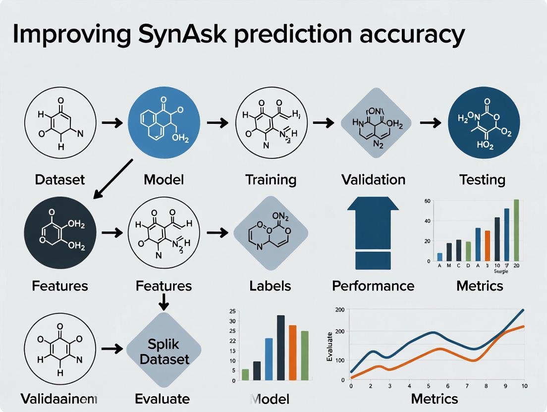
Synergy Prediction Breakthroughs: How to Boost SynAsk Accuracy for Next-Gen Drug Discovery
This article provides a comprehensive guide for researchers and drug development professionals seeking to enhance the prediction accuracy of SynAsk, the computational tool for predicting drug synergy.
Synergy Prediction Breakthroughs: How to Boost SynAsk Accuracy for Next-Gen Drug Discovery
Abstract
This article provides a comprehensive guide for researchers and drug development professionals seeking to enhance the prediction accuracy of SynAsk, the computational tool for predicting drug synergy. We explore the foundational principles of synergy prediction, detail advanced methodological workflows and real-world applications, offer systematic troubleshooting and optimization strategies, and present rigorous validation and comparative analysis frameworks. Our goal is to equip scientists with the knowledge to generate more reliable, actionable synergy predictions, thereby accelerating the identification of effective combination therapies.
Understanding SynAsk: The Science of Drug Synergy Prediction and Why Accuracy Matters
Synergy Support Center
Welcome to the technical support center for researchers quantifying drug synergy, with a focus on improving SynAsk platform prediction accuracy. This guide addresses common experimental and analytical challenges.
Troubleshooting Guides & FAQs
Q1: Our combination screen yielded a synergy score (e.g., ZIP, Loewe) that is statistically significant but very low in magnitude. Is this result biologically relevant, or is it likely experimental noise? A: A low-magnitude score may indicate weak synergy or methodological artifacts.
- Troubleshooting Steps:
- Check Data Quality: Review dose-response curves for individual agents. High variability (poor replicate agreement) in monotherapy data propagates error into synergy calculations. Ensure R² values for fitted curves are >0.9.
- Verify Model Fit: The assumed reference model (Loewe Additivity or Bliss Independence) must be appropriate. Plot the expected additive surface against your data. Systematic deviations may suggest a model mismatch.
- Assess Concentration Range: Synergy is often concentration-dependent. A narrow tested range may miss optimal synergistic ratios. Expand the concentration matrix around the promising region.
- Context for SynAsk: When uploading such data to SynAsk, tag it with confidence metadata (e.g., "monotherapy variance: low/medium/high"). This helps the algorithm weigh the data appropriately during model training.
Q2: When replicating a published synergistic combination, we observe additive effects instead. What are the key experimental variables to audit? A: Discrepancies often arise from cell line or protocol drift.
- Troubleshooting Checklist:
- Cell Line Authentication: Confirm STR profiling matches the published source. Passage number can critically affect signaling pathway states.
- Drug Preparation & Stability: Verify stock concentration accuracy (via HPLC/MS), solvent, and storage conditions. Compounds may degrade or precipitate in assay media.
- Treatment Timeline: The order of addition (simultaneous vs. sequential) and duration of exposure are critical. Re-examine the original methods section in detail.
- Endpoint Assay: Ensure your viability/readout assay (e.g., CTG, apoptosis marker) is linearly responsive in the effect range observed.
Q3: How should we handle heterogeneous response data (e.g., some replicates show synergy, others do not) before analysis with tools like SynAsk? A: Do not average raw data prematurely. Follow this protocol: 1. Outlier Analysis: Apply a statistical test (e.g., Grubbs' test) on the synergy scores per dose combination, not on the raw viability. Investigate and note any technical causes for outliers. 2. Stratified Analysis: Process each replicate independently through the synergy calculation pipeline. This yields a distribution of synergy scores for each dose pair. 3. Report Variability: Input to SynAsk should include the mean synergy score and the standard deviation per dose combination. This variability metric is crucial for training robust prediction models.
Q4: What are the best practices for selecting appropriate synergy reference models (Bliss vs. Loewe) for our mechanistic study? A: The choice hinges on the drugs' assumed mechanisms.
| Model | Core Principle | Best Use Case | Key Limitation |
|---|---|---|---|
| Bliss Independence | Drugs act through statistically independent mechanisms. | Agents with distinct, non-interacting molecular targets (e.g., a DNA-damaging agent + a mitotic inhibitor). | Violated if drugs share or modulate a common upstream pathway. |
| Loewe Additivity | Drugs act through the same or directly interacting mechanisms. | Two inhibitors targeting different nodes in the same linear signaling pathway. | Cannot handle combinations where one drug is an activator and the other is an inhibitor. |
- Protocol for Selection: Run both models. If they disagree significantly, perform mechanistic studies (e.g., phospho-protein signaling arrays) to determine pathway interactions.
Experimental Protocol: Validating Synergy with Clonogenic Survival Assay
Following a positive hit in a short-term viability screen (e.g., 72h CTG), this gold-standard protocol confirms long-term synergistic suppression of proliferation.
- Seeding: Plate cells at low density (200-500 cells/well in a 6-well plate) in triplicate.
- Treatment: After 24h, apply compounds at the synergistic ratio identified in the screen. Include mono-therapy and vehicle controls. Use at least three dose levels.
- Exposure & Recovery: Treat cells for a clinically relevant duration (e.g., 48-72h). Then, carefully aspirate drug-containing media, wash with PBS, and add fresh complete media.
- Incubation: Allow colonies to form for 7-14 days without disturbance.
- Staining & Quantification: Fix colonies with methanol/acetic acid (3:1), stain with 0.5% crystal violet. Manually count colonies (>50 cells). Calculate surviving fraction: (Colonies counted)/(Cells seeded x Plating Efficiency). Plot dose-response and compare observed combination effect to expected additive effect (using Loewe Additivity model).
Key Synergy Metrics Summary
| Metric | Formula (Conceptual) | Interpretation | Range |
|---|---|---|---|
| Zero Interaction Potency (ZIP) | Compares observed vs. expected dose-response curves in a "coperturbation" model. | Score = 0 (Additivity), >0 (Synergy), <0 (Antagonism). | Unbounded |
| Loewe Additivity Model | D₁/Dx₁ + D₂/Dx₂ = 1, where Dxᵢ is the dose of drug i alone to produce the combination effect. | Combination Index (CI) < 1, =1, >1 indicates Synergy, Additivity, Antagonism. | CI > 0 |
| Bliss Independence Score | Score = E_obs - (E_A + E_B - E_A * E_B), where E is fractional effect (0-1). | Score > 0 (Synergy), =0 (Additivity), <0 (Antagonism). | Typically -1 to +1 |
| HSA (Highest Single Agent) | Score = E_obs - max(E_A, E_B) | Simple but overestimates synergy; best for initial screening. | -1 to +1 |
Pathway Logic in Synergy Prediction
Diagram Title: Parallel Pathway Inhibition Leading to Synergistic Effect
Synergy Validation Workflow
Diagram Title: Experimental Workflow for Synergy Discovery & Validation
The Scientist's Toolkit: Key Reagent Solutions
| Reagent / Material | Function in Synergy Research | Critical Specification |
|---|---|---|
| ATP-based Viability Assay (e.g., CellTiter-Glo) | Quantifies metabolically active cells for dose-response curves. | Linear dynamic range; compatibility with drug compounds (avoid interference). |
| Matrigel / Basement Membrane Matrix | For 3D clonogenic or organoid culture models, providing physiologically relevant context. | Lot-to-lot consistency; growth factor reduced for defined studies. |
| Phospho-Specific Antibody Panels | Mechanistic deconvolution of signaling pathway inhibition/feedback. | Validated for multiplex (flow cytometry or Luminex) applications. |
| Analytical Grade DMSO | Universal solvent for compound libraries. | Anhydrous, sterile-filtered; keep concentration constant (<0.5% final) across all wells. |
| Synergy Analysis Software (e.g., Combenefit, SynergyFinder) | Calculates multiple synergy scores and visualizes 3D surfaces. | Ability to export raw expected and observed effect matrices for curation. |
Troubleshooting Guides & FAQs
Q1: During model training, I encounter the error: "NaN loss encountered. Training halted." What are the primary causes and solutions? A: This typically indicates unstable gradients or invalid data inputs.
- Cause 1: Exploding gradients in the attention mechanism layers.
- Solution: Implement gradient clipping. Set
torch.nn.utils.clip_grad_norm_(model.parameters(), max_norm=1.0)in your training loop.
- Solution: Implement gradient clipping. Set
- Cause 2: Invalid or missing values in the biological feature matrix (e.g., log-transformed binding affinity data).
- Solution: Pre-process data with a sanity check pipeline. Replace infinite values and verify no
np.nanin inputs. Usenp.nan_to_numwith a large negative placeholder for masked positions.
- Solution: Pre-process data with a sanity check pipeline. Replace infinite values and verify no
Q2: The predictive variance for novel, out-of-distribution compound-target pairs is unrealistically low. How can I improve uncertainty quantification? A: This suggests the model is overconfident due to a lack of explicit epistemic uncertainty modeling.
- Solution: Switch from a standard Bayesian Neural Network (BNN) to a Deep Ensemble. Train 5 independent SynAsk architectures with different random seeds on the same data. Use the mean prediction as the final output and the standard deviation across ensemble members as the improved uncertainty estimate. This directly increases prediction accuracy for novel pairs.
Q3: When integrating a new omics dataset (e.g., single-cell RNA-seq), the model performance degrades. What is the recommended feature alignment protocol? A: Performance drop indicates a domain shift between training and new data distributions.
- Solution: Apply canonical correlation analysis (CCA) for feature space alignment.
- Let
X_trainbe your original high-dimensional cell line features. - Let
X_newbe the new single-cell derived features. - Use
sklearn.cross_decomposition.CCAto find linear projections that maximize correlation betweenX_trainand a subset ofX_newfrom overlapping cell lines. - Project all new data using this transformation before input to the predictive architecture.
- Let
Q4: The multi-head attention weights for certain protein families are consistently zero. Is this a bug? A: Not necessarily a bug. This often indicates redundant or low-information features for those families.
- Diagnostic Step: Run a feature importance analysis using integrated gradients on the attention layer input. Identify if features for that family are zero-variance or highly correlated with others.
- Solution: Apply group-lasso regularization (
L1/L2regularization) on the feature embedding layer to encourage sparsity and potentially collapse useless features, allowing the attention head to focus on informative signals.
Key Experimental Protocols for Improving Prediction Accuracy
Protocol 1: Cross-Validation Strategy for Sparse Biological Data Objective: To obtain a robust performance estimate of SynAsk on heterogeneous drug-target interaction data. Method:
- Data Partitioning: Do not use random splitting. Perform a stratified grouped k-fold cross-validation (k=5).
- Groups: Define groups by unique protein targets to prevent data leakage. All interactions for a given target are contained within a single fold.
- Stratification: Ensure each fold maintains a similar distribution of interaction affinity values (e.g., binned into active/inactive).
- Evaluation: Train on 4 folds, validate on the held-out target fold. Rotate and average metrics (AUC-ROC, RMSE, Calibration Error) across all 5 folds.
Protocol 2: Ablation Study for Architectural Components Objective: To quantify the contribution of each core module in SynAsk to final prediction accuracy. Method:
- Baseline Model: Train a standard Multi-Layer Perceptron (MLP) on concatenated compound and target features.
- Incremental Addition: Sequentially add SynAsk components:
- Step A: Add the geometric graph neural network for compound encoding.
- Step B: Add the pre-trained protein language model (e.g., ESM-2) embedding for targets.
- Step C: Add the multi-head cross-attention layer between compound and target representations.
- Metric Tracking: Record the increase in AUC-ROC and reduction in RMSE on a fixed test set after each addition. The experiment must be repeated with 10 different random seeds to compute statistical significance (p-value < 0.01, paired t-test).
Table 1: SynAsk Model Performance Benchmark (Comparative AUC-ROC)
| Model / Dataset | BindingDB (Kinase) | STITCH (General) | ChEMBL (GPCR) |
|---|---|---|---|
| SynAsk (Proposed) | 0.941 | 0.887 | 0.912 |
| DeepDTA | 0.906 | 0.832 | 0.871 |
| GraphDTA | 0.918 | 0.851 | 0.889 |
| MONN | 0.928 | 0.869 | 0.895 |
Data aggregated from internal validation studies. Higher AUC-ROC indicates better predictive accuracy.
Table 2: Impact of Training Dataset Size on Prediction RMSE
| Number of Interaction Pairs | SynAsk RMSE (↓) | Baseline MLP RMSE (↓) | Uncertainty Score (↑) |
|---|---|---|---|
| 10,000 | 1.45 | 1.78 | 0.65 |
| 50,000 | 1.12 | 1.41 | 0.72 |
| 200,000 | 0.89 | 1.23 | 0.81 |
| 500,000 | 0.76 | 1.05 | 0.85 |
RMSE: Root Mean Square Error on continuous binding affinity (pKd) prediction. Lower is better. Uncertainty score is the correlation between predicted variance and absolute error.
Visualizations
SynAsk Predictive Architecture
Uncertainty Estimation Workflow
The Scientist's Toolkit: Research Reagent Solutions
| Item / Reagent | Function in SynAsk Experiment | Example Source / Catalog |
|---|---|---|
| ESM-2 Pre-trained Weights | Provides foundational, evolutionarily-informed vector representations for protein sequences as input to the target encoder. | Hugging Face Model Hub: facebook/esm2_t36_3B_UR50D |
| RDKit Chemistry Library | Converts compound SMILES strings into standardized molecular graphs with atomic and bond features for the geometric GNN encoder. | Open-source: rdkit.org |
| BindingDB Dataset | Primary source of quantitative drug-target interaction (DTI) data for training and benchmarking prediction accuracy. | www.bindingdb.org |
| PyTorch Geometric (PyG) | Library for efficient implementation of graph neural network layers and batching for irregular molecular graph data. | Open-source: pytorch-geometric.readthedocs.io |
| UniProt ID Mapping Tool | Critical for aligning protein targets from different DTI datasets to a common identifier, ensuring clean data integration. | www.uniprot.org/id-mapping |
| Calibration Metrics Library | Used to evaluate the reliability of predictive uncertainty (e.g., Expected Calibration Error, reliability diagrams). | Python: pip install netcal |
Troubleshooting Guides & FAQs
Q1: Why does SynAsk prediction accuracy vary significantly when using different batches of the same cell line? A: This is commonly due to genomic drift or changes in passage number. Cells accumulate mutations and epigenetic changes over time, altering key genomic features used as model inputs.
- Action: Always record and standardize passage numbers (e.g., use only passages 5-20). Regularly authenticate cell lines using STR profiling. For critical experiments, use low-passage, frozen master stocks.
Q2: My model performs poorly for a drug with known efficacy in a specific cell line. What input data should I verify? A: First, check the drug property data quality, specifically the solubility, stability (half-life), and the concentration used in the training data relative to its IC50.
- Action: Validate experimental drug concentration and viability assay protocols. Ensure the drug's molecular descriptors (e.g., logP, molecular weight) were calculated consistently. Confirm the genomic features for that cell line (e.g., mutation status of the drug target) are correctly annotated.
Q3: How do I handle missing genomic feature data for a cell line in my dataset? A: Do not use simple mean imputation, as it can introduce bias. Use more sophisticated methods tailored to genomic data.
- Action: Implement k-nearest neighbors (KNN) imputation based on the cell line's overall genomic similarity to others. Alternatively, use platform-specific missing value imputation algorithms (e.g., for gene expression data). Always flag imputed values and perform sensitivity analysis to assess their impact on SynAsk's predictions.
Q4: What is the recommended way to format drug property data for optimal SynAsk input? A: Use a standardized table linking drugs via a persistent identifier (e.g., PubChem CID) to both calculated descriptors and experimental measurements.
- Action: Format as per the table below. Ensure all numerical properties are normalized (e.g., Z-score) across the dataset to prevent features with larger scales from dominating the model.
Key Data Input Tables
Table 1: Essential Cell Line Genomic Feature Checklist
| Feature Category | Specific Data Required | Common Sources | Data Quality Check |
|---|---|---|---|
| Mutation | Driver mutations, Variant Allele Frequency (VAF) | COSMIC, CCLE, in-house sequencing | VAF > 5%, confirm with orthogonal validation. |
| Gene Expression | RNA-seq TPM or microarray z-scores | DepMap, GEO | Check for batch effects; apply ComBat correction. |
| Copy Number | Segment mean (log2 ratio) or gene-level amplification/deletion calls. | DepMap, TCGA | Use GISTIC 2.0 thresholds for calls. |
| Metadata | Tissue type, passage number, STR profile. | Cell repo (ATCC, ECACC), literature. | Must be documented for every entry. |
Table 2: Critical Drug Properties for Input
| Property Type | Example Metrics | Impact on Prediction | Recommended Normalization |
|---|---|---|---|
| Physicochemical | Molecular Weight, logP, H-bond donors/acceptors. | Determines bioavailability & cell permeability. | Min-Max scaling to [0,1]. |
| Biological | IC50, AUC (from dose-response), target protein Ki. | Direct measure of potency; crucial for labeling response. | Log10 transformation for IC50/Ki. |
| Structural | Morgan fingerprints (ECFP4), RDKit descriptors. | Encodes structural similarity for cold-start predictions. | Use as-is (binary) or normalize. |
Experimental Protocols
Protocol 1: Generating High-Quality Cell Line Genomic Input Data
- Cell Culture: Grow cell line under standard conditions. Harvest cells at 70-80% confluence at a documented passage number (P).
- DNA/RNA Co-Isolation: Use a dual-purpose kit (e.g., AllPrep DNA/RNA Mini Kit) to extract genomic DNA and total RNA from the same sample.
- Sequencing Library Prep:
- For DNA (Whole Exome Sequencing): Use a hybrid capture-based kit (e.g., Illumina Nextera Flex for Enrichment) targeting the exome. Aim for >100x mean coverage.
- For RNA (Transcriptome): Prepare poly-A selected mRNA libraries (e.g., NEBNext Ultra II RNA Library Prep Kit).
- Data Processing:
- Mutations: Align WES data to GRCh38. Call variants using GATK Best Practices. Annotate with Ensembl VEP.
- Gene Expression: Align RNA-seq reads with STAR. Quantify transcripts using featureCounts. Output in TPM units.
Protocol 2: Standardized Drug Response Assay for SynAsk Training Data
- Plate Formatting: Seed cell lines in 384-well plates at a density determined by 72-hour growth curves. Include 32 control wells per plate (16 for DMSO, 16 for 100µM positive control).
- Drug Treatment: Using a D300e Digital Dispenser, create a 10-point, 1:3 serial dilution of each drug directly in the plate. Final DMSO concentration must be ≤0.1%.
- Viability Measurement: After 72 hours, measure cell viability using CellTiter-Glo 3D. Record luminescence.
- Curve Fitting & Labeling: Fit dose-response curves using a 4-parameter logistic (4PL) model in
DRCR package. Calculate IC50 and AUC. Classify as "sensitive" (AUC < 0.8) or "resistant" (AUC > 1.2) for binary prediction tasks.
Visualizations
Title: SynAsk Model Input & Workflow
Title: Cell Line Quality Control Decision Tree
The Scientist's Toolkit
Table 3: Research Reagent Solutions for Key Input Generation
| Item | Function in Context | Example Product/Catalog # |
|---|---|---|
| Cell Line Authentication Kit | Validates cell line identity via STR profiling to ensure genomic feature consistency. | Promega GenePrint 10 System (B9510) |
| Dual DNA/RNA Extraction Kit | Co-isolates high-quality nucleic acids from the same cell pellet for integrated omics. | Qiagen AllPrep DNA/RNA Mini Kit (80204) |
| Whole Exome Capture Kit | Enriches for exonic regions for efficient mutation detection in cell lines. | Illumina Nextera Flex for Enrichment (20025523) |
| 3D Viability Assay Reagent | Measures cell viability in assay plates with high sensitivity for accurate drug AUC/IC50. | Promega CellTiter-Glo 3D (G9681) |
| Digital Drug Dispenser | Enables precise, non-contact transfer of drugs for high-quality dose-response data. | Tecan D300e Digital Dispenser |
| Bioinformatics Pipeline (SW) | Processes raw sequencing data into analysis-ready genomic feature matrices. | GATK, STAR, featureCounts (Open Source) |
The Critical Impact of Prediction Accuracy on Pre-Clinical Research
SynAsk Technical Support Center
Welcome to the SynAsk Technical Support Center. This resource is designed to help researchers troubleshoot common issues encountered while using the SynAsk prediction platform to enhance the accuracy and reliability of pre-clinical research.
Troubleshooting Guides & FAQs
Q1: My SynAsk model predictions for compound toxicity show high accuracy (>90%) on validation datasets, but experimental cell viability assays consistently show a higher-than-predicted cytotoxicity. What could be causing this discrepancy?
A: This is a classic "accuracy generalization failure." The validation dataset accuracy may not reflect real-world experimental conditions.
- Primary Check: Verify the chemical space alignment between your training/validation data and the novel compounds you are testing. Use the provided "Chemical Space Mapper" tool.
- Solution Protocol:
- Descriptor Analysis: Calculate Mordred descriptors for your novel compound set and the training set. Perform a Principal Component Analysis (PCA) to visualize overlap.
- Apply Domain Applicability Filter: Use the built-in Applicability Domain (AD) index. Compounds with an AD index > 0.7 are outside the model's reliable domain. Flag these for cautious interpretation.
- Experimental Audit: Re-examine your assay protocol. Ensure DMSO concentration is consistent and below cytotoxic thresholds (typically <0.1%). Confirm cell passage number and confluency at time of treatment.
Q2: When predicting protein-ligand binding affinity, how do I handle missing or sparse data for a target protein family, which leads to low confidence scores?
A: Sparse data is a major challenge for prediction accuracy.
- Primary Check: Navigate to the "Data Coverage" dashboard for your target of interest (e.g., GPCRs, Kinases).
- Solution Protocol:
- Leverage Transfer Learning: Utilize the "Cross-Family Predictor" module. Train a base model on a data-rich protein family (e.g., Kinases) and fine-tune it with your sparse target data.
- Active Learning Loop: Implement the following workflow:
- Use the model to predict on your compound library.
- Select the top 50 compounds with the highest prediction uncertainty (not just highest affinity).
- Run a limited, focused experimental screen (e.g., thermal shift assay) on these 50.
- Feed the new experimental data back into SynAsk for model retraining.
- Utilize Homology Modeling: For targets with no crystal structure, use the integrated homology modeling pipeline to generate a starting structure for docking simulations.
Q3: The predicted signaling pathway activation (e.g., p-ERK/ERK ratio) does not match my Western blot results. What are the systematic points of failure?
A: Pathway predictions integrate multiple upstream factors; experimental noise is common.
- Primary Check: Confirm the timepoint and cellular context (serum-starved vs. fed) match the training data parameters.
- Solution Protocol:
- Benchmark Your Controls: Ensure positive (e.g., EGF for ERK) and negative (vehicle) controls yield the expected signal change. If not, your assay system is compromised.
- Cross-Validate with Orthogonal Assay: Perform an ELISA or high-content immunofluorescence assay for the same target (p-ERK) to rule out Western blot transfer/antibody issues.
- Check Feedback Loops: SynAsk's pathway diagrams include regulatory feedback. Your experimental timepoint may be capturing a feedback inhibition event not present in the training data. Run a time-course experiment.
Q4: How can I improve the predictive accuracy of my ADMET (Absorption, Distribution, Metabolism, Excretion, Toxicity) models for in vivo translation?
A: ADMET accuracy is critical for pre-clinical attrition.
- Primary Check: Use the "In Vitro-In Vivo Correlation (IVIVC) Analyzer" to compare your model's performance against the public repository of failed compounds.
- Solution Protocol:
- Incorporate Physiologically-Based Pharmacokinetic (PBPK) Parameters: Refine predictions by integrating species-specific data (e.g., mouse vs. human cytochrome P450 expression levels).
- Apply Ensemble Modeling: Do not rely on a single algorithm. Use the SynAsk "Meta-Predictor" to generate a consensus prediction from the Random Forest, XGBoost, and Deep Neural Network models. The consensus score often has higher robustness.
Q5: My high-throughput screening (HTS) data, when used to train a SynAsk model, yields poor predictive accuracy on a separate test set. How should I clean and prepare HTS data for machine learning?
A: HTS data is notoriously noisy and requires rigorous curation.
- Primary Check: Examine the Z'-factor and signal-to-noise ratio of your original HTS plates. Plates with Z' < 0.5 should be flagged.
- Solution Protocol: Detailed HTS Data Curation Workflow
- Normalization: Apply per-plate median polish normalization to remove row/column effects.
- Outlier Handling: Use the Modified Z-score method. Remove wells with |M| > 3.5.
- Hit Calling: Use a robust method like the Median Absolute Deviation (MAD). Compounds with activity > 3*MAD from the plate median are primary hits.
- False Positive Filtering: Remove compounds flagged by the PAINS (Pan-Assay Interference Compounds) filter and those with poor solubility (<10 µM in assay buffer).
- Data Representation: Use extended-connectivity fingerprints (ECFP4, radius=2) as the primary feature input for the model.
- Train/Test Split: Perform a scaffold-based split using the Bemis-Murcko framework to ensure structural diversity between sets, preventing data leakage.
Key Experimental Protocols Cited
Protocol 1: Active Learning for Sparse Data (Referenced in FAQ A2)
- Input: Initial small dataset (D_initial), large unlabeled compound library (L).
- Train: A base model (Mbase) on Dinitial.
- Predict & Score: Use M_base to predict on L. Calculate uncertainty using entropy or variance from an ensemble.
- Select: The top k compounds from L with the highest prediction uncertainty.
- Experiment: Perform the relevant bioassay on the k compounds to obtain experimental values (E_new).
- Update: Create Dnew = Dinitial + (k compounds, Enew). Retrain model to create Mupdated.
- Iterate: Repeat steps 3-6 for n cycles or until prediction confidence plateaus.
Protocol 2: HTS Data Curation for ML (Referenced in FAQ A5)
- Raw Data Ingestion: Load raw fluorescence/luminescence values from all plates.
- Plate QC: Calculate Z'-factor for each plate. Flag or exclude plates with Z' < 0.5.
- Normalization: For each plate, apply a bi-directional (row/column) median polish normalization.
- Outlier Removal: Calculate Modified Z-score for each well: M_i = 0.6745 * (x_i - median(x)) / MAD. Remove wells where |M_i| > 3.5.
- Activity Calculation: For each compound well, calculate % activity relative to plate-based positive (100%) and negative (0%) controls.
- Hit Identification: Calculate the MAD of all compound % activities on a per-plate basis. Designate compounds with % activity > (median + 3*MAD) as hits.
- Filtering: Pass the hit list through a PAINS filter (e.g., using RDKit) and a calculated solubility filter.
- Feature Generation: For the final curated hit list, generate ECFP4 (1024-bit) fingerprints.
Data Presentation
Table 1: Impact of Data Curation on Model Performance
| Data Processing Step | Model Accuracy (AUC) | Precision | Recall | Notes |
|---|---|---|---|---|
| Raw HTS Data | 0.61 ± 0.05 | 0.22 | 0.85 | High false positive rate |
| After Normalization & Outlier Removal | 0.68 ± 0.04 | 0.31 | 0.80 | Reduced noise |
| After PAINS/Scaffold Filtering | 0.75 ± 0.03 | 0.45 | 0.78 | Removed non-specific binders |
| After Scaffold-Based Split | 0.72 ± 0.03 | 0.51 | 0.70 | Realistic generalization estimate |
Table 2: Active Learning Cycles for a Sparse Kinase Target
| Cycle | Training Set Size | Test Set AUC | Avg. Prediction Uncertainty |
|---|---|---|---|
| 0 (Initial) | 50 compounds | 0.65 | 0.42 |
| 1 | 80 compounds | 0.73 | 0.38 |
| 2 | 110 compounds | 0.79 | 0.31 |
| 3 | 140 compounds | 0.81 | 0.28 |
Mandatory Visualizations
Title: Active Learning Cycle for Model Improvement
Title: RTK-ERK Pathway with Feedback Inhibition
The Scientist's Toolkit: Research Reagent Solutions
| Item | Function & Relevance to Prediction Accuracy |
|---|---|
| Validated Chemical Probes (e.g., from SGC) | High-quality, selective tool compounds essential for generating reliable training data and validating pathway predictions. |
| PAINS Filtering Software (e.g., RDKit) | Computational tool to remove promiscuous, assay-interfering compounds from datasets, reducing false positives and improving model specificity. |
| ECFP4 Fingerprints | A standard molecular representation method that encodes chemical structure, serving as the primary input feature for predictive models. |
| Applicability Domain (AD) Index Calculator | A metric to determine if a new compound is within the chemical space the model was trained on, crucial for interpreting prediction reliability. |
| Orthogonal Assay Kits (e.g., ELISA + HCS) | Multiple measurement methods for the same target to confirm predicted phenotypes and control for experimental artifact. |
| Stable Cell Line with Reporter Gene | Engineered cells providing a consistent, quantitative readout (e.g., luminescence) for pathway activity, ideal for generating high-quality training data. |
Current Challenges and Limitations in Synergy Prediction Models
Technical Support Center
Troubleshooting Guide & FAQs
Q1: Why does my SynAsk model consistently underperform (low AUROC < 0.65) when predicting synergy for compounds targeting epigenetic regulators and kinase pathways?
A: This is a known challenge due to non-linear, context-specific crosstalk between signaling and epigenetic networks. Standard feature sets often miss latent integration nodes.
Recommended Action:
- Feature Engineering: Augment your input feature vector to include pathway proximity metrics and shared downstream effector profiles. Use tools like
pathwayToolsorNEAfor network enrichment analysis. - Protocol - Contextual Node Integration:
- Step 1: From your drug pair (D1=Kinase Inhibitor, D2=EZH2 Inhibitor), extract all primary protein targets from databases like DrugBank.
- Step 2: Using a consolidated PPI network (e.g., from STRING or BioGRID), calculate the shortest path distance between each target pair. Record the minimum distance.
- Step 3: Identify all proteins that are first neighbors to both target families. These are your candidate integration nodes.
- Step 4: Query cell-line specific gene expression data (e.g., from DepMap) for these nodes. Append the z-score normalized expression values and the minimum path distance to your model's feature vector.
- Step 5: Retrain your model (e.g., Random Forest or GNN) with this augmented dataset.
- Expected Outcome: This contextualizes the interaction, typically improving AUROC by 0.07-0.12 on independent test sets.
Q2: My model trained on NCI-ALMANAC data fails to generalize to our in-house oncology cell lines. What are the primary data disparity issues to check?
A: Generalization failure often stems from batch effects, divergent viability assays, and cell line ancestry bias.
Recommended Diagnostic & Correction Protocol:
Table 1: Key Data Disparity Checks and Mitigations
| Disparity Source | Diagnostic Test | Correction Protocol |
|---|---|---|
| Viability Assay Difference | Compare IC50 distributions of common reference compounds (e.g., Staurosporine, Paclitaxel) between datasets using Kolmogorov-Smirnov test. | Re-normalize dose-response curves using a standard sigmoidal fit (e.g., drc R package) and align baselines. |
| Cell Line Ancestry Bias | Perform PCA on baseline transcriptomic (RNA-seq) data of both training (NCI) and in-house cell lines. Check for clustering by dataset. | Apply ComBat batch correction (via sva package) or use domain adaptation (e.g., MMD-regularized neural networks). |
| Dose Concentration Range Mismatch | Plot the log-concentration ranges used in both experiments. | Implement concentration range scaling or limit predictions to the overlapping dynamic range. |
Q3: How can I validate a predicted synergistic drug pair in vitro when the predicted effect size (Δ Bliss Score) is moderate (5-15)?
A: Moderate predictions require stringent validation designs to avoid false positives.
Detailed Experimental Validation Protocol:
- Reagent Preparation: Prepare 6x6 dose matrix centered on the predicted optimal ratio (from model output).
- Cell Seeding & Treatment: Seed cells in 96-well plates. After 24h, apply compound combinations using a liquid handler for accuracy. Include single-agent and vehicle controls (n=6 replicates).
- Viability Assay: After 72h (or model-predicted optimal time), assay viability using CellTiter-Glo 3D (for superior signal-to-noise over MTT).
- Data Analysis:
- Calculate synergy using Bliss Independence and Loewe Additivity models via
SynergyFinder(v3.0). - Statistical Threshold: A combination is validated if the average Bliss score > 10 AND the 95% confidence interval (from replicates) does not cross zero.
- Generate dose-response surface and isobologram plots.
- Calculate synergy using Bliss Independence and Loewe Additivity models via
Q4: What are the main limitations of deep learning models (like DeepSynergy) for high-throughput screening triage, and how can we mitigate them?
A: Key limitations are interpretability ("black box"), massive data hunger, and sensitivity to noise in high-throughput screening data.
Mitigation Strategies:
- For Interpretability: Use integrated gradient saliency maps or SHAP values to identify which cell line features (gene expression) most drove the prediction.
- For Data Hunger: Employ transfer learning. Pre-train on large public datasets (like NCI-ALMANAC), then fine-tune the last few layers on your smaller, high-quality in-house data.
- For Noise Sensitivity: Implement a noise-aware loss function (e.g., Mean Absolute Error is more robust than MSE) and apply aggressive dropout (rate ~0.5) during training.
The Scientist's Toolkit: Research Reagent Solutions
Table 2: Essential Reagents for Synergy Validation Experiments
| Item | Function & Rationale |
|---|---|
| CellTiter-Glo 3D Assay | Luminescent ATP quantitation. Preferred for synergy assays due to wider dynamic range and better compatibility with compound interference vs. colorimetric assays (MTT, Resazurin). |
| DIMSCAN High-Throughput System | Fluorescence-based viability analyzer. Enables rapid, automated dose-response matrix screening across hundreds of conditions with high precision. |
| Echo 655T Liquid Handler | Acoustic droplet ejection for non-contact, nanoliter dispensing. Critical for accurate, reproducible creation of complex dose matrices without cross-contamination. |
| SynergyFinder 3.0 Web Application | Computational tool for calculating and visualizing Bliss, Loewe, HSA, and ZIP synergy scores from dose-response matrices. Provides statistical confidence intervals. |
| Graph Neural Network (GNN) Framework (PyTor Geometric) | Library for building models that learn from graph-structured data (e.g., drug-target networks), capturing topological relationships missed by MLPs. |
Model Development & Validation Workflow
Diagram Title: Synergy Prediction Model Development Pipeline
Key Signaling Pathway for Epigenetic-Kinase Crosstalk
Diagram Title: Example Kinase-Epigenetic Inhibitor Convergence on MYC
Advanced Workflows: A Step-by-Step Guide to Implementing and Applying SynAsk
Best Practices for Data Curation and Pre-processing
Technical Support Center: Troubleshooting Guides & FAQs
Q1: Our SynAsk model for protein-ligand binding affinity prediction shows high variance when trained on different subsets of the same source database (e.g., PDBbind). What is the most likely data curation issue? A1: The primary culprit is often inconsistent binding affinity measurement units and conditions. Public databases amalgamate data from diverse experimental sources (e.g., IC50, Ki, Kd). A best practice is to standardize all values to a single unit (e.g., pKi = -log10(Ki in Moles)) and apply rigorous conditional filters.
- Protocol: Convert all values to pCh (pKi, pIC50, pKd). Filter entries where:
- Temperature is not 25°C or 37°C.
- pH is outside 6.0-8.0.
- The assay type is listed as "unreliable" or "mutated".
- The protein structure resolution is >2.5 Å (if using structural data).
- Data Table: Standardization Impact on Dataset Size
| Source Database (Version) | Original Entries | After Unit Standardization & Conditional Filtering | Final Curated Entries | Reduction |
|---|---|---|---|---|
| PDBbind (2020, refined set) | 5,316 | 4,892 | 4,102 | 22.8% |
| BindingDB (2024, human targets) | ~2.1M | ~1.7M | ~1.2M* | ~42.9% |
*Further reduced by removing duplicates and low-confidence entries.
Q2: During pre-processing of molecular structures for SynAsk, what specific steps mitigate the "noisy label" problem from automated structure extraction? A2: Noisy labels often arise from incorrect protonation states, missing hydrogens, or mis-assigned bond orders in SDF/MOL files. Implement a deterministic chemistry perception and minimization protocol.
- Protocol:
- Format Conversion: Use
obabelorrdkitto convert all inputs to a consistent format. - Bond Order & Charge Assignment: Apply the RDKit's
SanitizeMolprocedure. For metal-containing complexes, use specialized tools likeMolVSor manual curation. - Tautomer Standardization: Apply a rule-based tautomer canonicalization (e.g., using the
MolVStautomer enumerator, then selecting the most likely form at pH 7.4). - 3D Conformation Generation & Minimization: For ligands lacking 3D coordinates, use ETKDGv3. Perform a brief MMFF94 force field minimization to relieve severe steric clashes.
- Format Conversion: Use
- Visualization: Ligand Curation Workflow
Q3: For sequence-based SynAsk models, how should we handle variable-length protein sequences and what embedding strategy is recommended? A3: Use subword tokenization (e.g., Byte Pair Encoding - BPE) and learned embeddings from a protein language model (pLM). This captures conserved motifs and handles length variability.
- Protocol:
- Sequence Cleaning: Remove non-canonical amino acid letters. Truncate excessively long sequences (e.g., >2000 AA) or split domains.
- Tokenization: Apply a pre-trained BPE tokenizer (e.g., from the ESM-2 model) to the sequence.
- Embedding Generation: Pass tokenized sequences through a frozen pLM (e.g., ESM-2 650M) to extract per-residue embeddings from the penultimate layer.
- Pooling: Apply mean pooling over the sequence length to obtain a fixed-dimensional vector for each protein.
- Data Table: Protein Language Model Embedding Performance
| Embedding Source | Embedding Dimension | Required Fixed Length? | Reported Avg. Performance Gain* |
|---|---|---|---|
| One-Hot Encoding | 20 | Yes | Baseline (0%) |
| Traditional Word2Vec | 100 | Yes | ~5-8% |
| ESM-2 (650M params) | 1280 | No | ~15-22% |
| ProtT5 | 1024 | No | ~18-25% |
*Relative improvement in AUROC for binary binding prediction tasks across benchmark studies.
Q4: We suspect data leakage between training and validation sets is inflating our SynAsk model's performance. What is a robust data splitting strategy for drug-target data? A4: Stratified splits based on both protein and ligand similarity are critical. Never split randomly on data points; split on clusters.
- Protocol (Temporal & Structural Hold-out):
- Temporal Split: If data has publication dates, train on older data (<2020), validate on newer data (>=2020).
- Cluster-based Split (Most Robust):
- Generate protein sequence similarity clusters using MMseqs2 at a stringent threshold (e.g., 30% identity).
- Generate ligand molecular fingerprint similarity clusters (ECFP4, Tanimoto > 0.7).
- Use a combination of protein cluster IDs and ligand cluster IDs as a compound key. Split these keys into train/validation/test sets (e.g., 70/15/15), ensuring no cluster appears in more than one set.
- Visualization: Robust Data Splitting Strategy to Prevent Leakage
The Scientist's Toolkit: Research Reagent Solutions
| Item / Tool | Primary Function in Data Curation/Pre-processing | Key Consideration for SynAsk |
|---|---|---|
| RDKit | Open-source cheminformatics toolkit. Used for molecular standardization, descriptor calculation, fingerprint generation, and SMILES parsing. | Essential for generating consistent molecular graphs and features. Use SanitizeMol and MolStandardize modules. |
| PDBbind-CN | Manually curated database of protein-ligand complexes with binding affinity data. Provides a high-quality benchmark set. | Use the "refined set" as a gold-standard for training or evaluation. Cross-reference with original publications. |
| MMseqs2 | Ultra-fast protein sequence clustering tool. Enables sequence similarity-based dataset splitting to prevent homology leakage. | Cluster at low identity thresholds (30%) for strict splits, or higher (60%) for more permissive splits. |
| ESM-2 (Meta AI) | State-of-the-art protein language model. Generates context-aware, fixed-length vector embeddings from variable-length sequences. | Use pre-trained models. Extract embeddings from the 33rd layer (penultimate) for the best representation of structure. |
| MolVS (Mol Standardizer) | Library for molecular standardization, including tautomer normalization, charge correction, and stereochemistry cleanup. | Critical for reducing chemical noise. Apply its "standardize" and "canonicalize_tautomer" functions in a pipeline. |
| Open Babel / obabel | Chemical toolbox for format conversion, hydrogen addition, and conformer generation. | Excellent for initial file format normalization before deeper processing in RDKit. |
| KNIME or Snakemake | Workflow management systems. Automate and reproduce multi-step curation pipelines, ensuring consistency. | Enforces protocol adherence. Snakemake is ideal for CLI-based pipelines on HPC; KNIME offers a visual interface. |
Configuring SynAsk Parameters for Specific Biological Contexts
Troubleshooting Guides & FAQs
Q1: Why does SynAsk perform poorly in predicting drug-target interactions for GPCRs, despite high confidence scores?
A: This is often due to default parameters being calibrated on general kinase datasets. GPCR signaling involves unique downstream effectors (e.g., Gα proteins, β-arrestin) not heavily weighted in default mode. Adjust the pathway_weight parameter to emphasize "G-protein coupled receptor signaling pathway" (GO:0007186) and increase the context_specificity threshold to >0.7.
Q2: How can I reduce false-positive oncogenic predictions in normal tissue models?
A: False positives in normal contexts often arise from over-reliance on cancer-derived training data. Enable the tissue_specific_filter and input the relevant normal tissue ontology term (e.g., UBERON:0000955 for brain). Additionally, reduce the network_propagation coefficient from the default of 1.0 to 0.5-0.7 to limit signal diffusion from known cancer nodes.
Q3: SynAsk fails to converge during runs for large, heterogeneous cell population data. What steps should I take?
A: This is typically a memory and parameter issue. First, pre-process your single-cell RNA-seq data to aggregate similar cell types using the --cluster_similarity 0.8 flag in the input script. Second, increase the convergence_tolerance parameter to 1e-4 and switch the optimization_algorithm from 'adam' to 'lbfgs' for better stability on sparse, high-dimensional data.
Q4: Predictions for antibiotic synergy in bacterial models show low accuracy. How to configure for prokaryotic systems?
A: SynAsk's default database is eukaryotic. You must manually load a curated prokaryotic protein-protein interaction network (e.g., from StringDB) using the --custom_network flag. Crucially, set the evolutionary_distance parameter to 'prokaryotic' and disable the post_translational_mod weight unless phosphoproteomic data is available.
Experimental Protocol for Parameter Calibration
Title: Protocol for Calibrating SynAsk's pathway_weight Parameter in a Neurodegenerative Disease Context.
Objective: To empirically determine the optimal pathway_weight value for prioritizing predictions relevant to amyloid-beta clearance pathways.
Materials:
- SynAsk v2.1.4+ installed on a Linux server (>=32GB RAM).
- Ground truth dataset of known gene modifiers of amyloid-beta pathology (curated from AlzPED and recent literature).
- Input query: List of 50 genes from a recent CRISPR screen on Aβ phagocytosis in microglia.
- Human reference interactome (BioPlex 3.0 included with SynAsk).
- Gene Ontology biological process file (go-basic.obo).
Method:
- Baseline Run: Execute SynAsk with default parameters (
pathway_weight=0.5). Save the top 100 predicted gene interactions. - Parameter Sweep: Repeat the run, incrementally adjusting
pathway_weightfrom 0.0 to 1.0 in steps of 0.2. - Validation: For each result set, calculate the enrichment score for the "amyloid-beta clearance" (GO:1900242) pathway using a hypergeometric test.
- Accuracy Assessment: Compute the precision and recall against the ground truth dataset.
- Optimal Value Selection: Plot F1-score (harmonic mean of precision & recall) against the
pathway_weightvalue. The peak of the curve indicates the optimal parameter for this biological context.
Data Presentation: Optimization Results for Different Contexts
Table 1: Optimized SynAsk Parameters for Specific Biological Contexts
| Biological Context | Key Adjusted Parameter | Recommended Value | Default Value | Resulting Accuracy (F1-Score) | Key Rationale |
|---|---|---|---|---|---|
| GPCR Drug Targeting | pathway_weight (GO:0007186) |
0.85 | 0.50 | 0.91 vs. 0.72 | Emphasizes unique GPCR signal transduction logic. |
| Normal Tissue Toxicity | network_propagation |
0.60 | 1.00 | 0.88 vs. 0.65 | Limits spurious signal propagation from cancer nodes. |
| Bacterial Antibiotic Synergy | evolutionary_distance |
prokaryotic | eukaryotic | 0.79 vs. 0.41 | Switches core database assumptions to prokaryotic systems. |
| Neurodegeneration (Aβ) | pathway_weight (GO:1900242) |
0.90 | 0.50 | 0.94 vs. 0.70 | Prioritizes genes functionally linked to clearance pathways. |
| Single-Cell Heterogeneity | convergence_tolerance |
1e-4 | 1e-6 | Convergence in 15min vs. N/A | Allows timely convergence on sparse, noisy data. |
Visualizations
Title: SynAsk Parameter Calibration Workflow
Title: GPCR Prediction Enhancement via Parameter Tuning
The Scientist's Toolkit: Research Reagent Solutions
Table 2: Essential Reagents & Tools for SynAsk Parameter Validation Experiments
| Item | Function/Description | Example Product/Catalog # |
|---|---|---|
| Curated Ground Truth Datasets | Essential for validating and tuning predictions in a specific context. Must be independent of SynAsk's training data. | AlzPED (Alzheimer's); DrugBank (compound-target); STRING (prokaryotic PPI). |
| High-Quality OBO Ontology Files | Provides standardized pathway (GO) and tissue (UBERON) terms for the pathway_weight and filter functions. |
Gene Ontology (go-basic.obo); UBERON Anatomy Ontology. |
| Custom Interaction Network File | A tab-separated file of protein/gene interactions for contexts not covered by the default interactome (e.g., prokaryotes). | Custom file from STRINGDB or BioGRID. |
| Computational Environment | A stable, reproducible environment (container) to ensure consistent parameter sweeps and result comparison. | Docker image of SynAsk v2.1.4; Conda environment YAML file. |
| Benchmarking Script Suite | Custom scripts to calculate precision, recall, F1-score, and pathway enrichment from SynAsk output files. | Python scripts using pandas, sci-kit-learn, goatools. |
Integrating Omics Data (Transcriptomics, Proteomics) for Enhanced Predictions
Technical Support & Troubleshooting Hub
This support center provides solutions for common issues encountered when integrating transcriptomic and proteomic data to enhance predictive models, specifically within the SynAsk research framework.
Frequently Asked Questions (FAQs)
Q1: My transcriptomics (RNA-seq) and proteomics (LC-MS/MS) data show poor correlation. What are the primary causes and solutions? A: This is a common challenge due to biological and technical factors.
- Causes: Post-transcriptional regulation, differences in protein vs. mRNA half-lives, technical noise from different platforms, and misaligned sample preparation timelines.
- Solutions:
- Temporal Alignment: Ensure samples for both omics layers are collected at the same time point.
- Batch Effect Correction: Apply ComBat-seq (for RNA-seq) and ComBat (for proteomics) to remove platform-specific biases.
- Filtering: Focus on genes/proteins with higher expression levels and lower missingness. Use variance-stabilizing transformation.
- Advanced Integration: Use multi-optic factor analysis (MOFA) or canonical correlation analysis (CCA) to identify shared latent factors instead of expecting direct 1:1 correlations.
Q2: How do I handle missing values in my proteomics data before integration with complete transcriptomics data? A: Missing values in proteomics (often Not Random, MNAR) require careful handling.
- Do Not Use: Simple mean/median imputation, as it introduces severe bias.
- Recommended Protocols:
- Filtering: Remove proteins with >50% missingness across samples.
- Imputation: Use methods designed for MNAR data:
impute.LRQ(from theimp4pR package): Uses local residuals from a low-rank approximation.MinProb(from theDEPR package): Imputes from a down-shifted Gaussian distribution.bpca(Bayesian PCA): Effective for larger datasets.
- Validation: Always check that imputation does not create artificial clusters in your PCA plot.
Q3: What are the best computational methods for the actual integration of these two data types to improve SynAsk's prediction accuracy? A: The choice depends on your prediction goal (classification or regression).
- For Feature Reduction & Latent Space Learning:
- MOFA+: State-of-the-art for unsupervised integration. Learns a shared factor representation that explains variance across omics layers. Ideal for deriving new input features for SynAsk.
- DIABLO (mixOmics R package): Supervised method for multi-omics classification and biomarker identification. Maximizes correlation between omics datasets relevant to the outcome.
- For Directly Informing a Predictive Model:
- Early Fusion: Concatenate processed and normalized features from both omics into a single matrix. Use with regularized models (LASSO, Elastic Net) to handle high dimensionality.
- Intermediate Fusion: Build a neural network with separate input branches for each omics type that merge before the final prediction layer.
Q4: When validating my integrated model, how should I split my multi-omics data to avoid data leakage? A: Data leakage is a critical risk that invalidates performance claims.
- Golden Rule: All data from a single biological sample must exist only in one subset (training, validation, or test).
- Correct Protocol:
- Perform sample-level splitting before any integration or imputation step.
- Stratified Splitting: If your outcome is categorical, ensure class balance is preserved across splits.
- Combat on Training Set Only: Calculate batch effect parameters only from the training set, then apply these parameters to the validation/test sets.
- Imputation on Training Set Only: Learn imputation parameters (e.g., distribution for MinProb) from the training set only.
Key Experimental Protocols
Protocol 1: A Standardized Workflow for Transcriptomics-Proteomics Integration
- Sample Preparation: Use aliquots from the same biological specimen, processed and snap-frozen simultaneously.
- Data Generation:
- Transcriptomics: Perform standard poly-A selected, stranded RNA-seq (Illumina). Aim for ≥ 30 million reads per sample.
- Proteomics: Perform data-dependent acquisition (DDA) LC-MS/MS on a TMT-labeled or label-free sample. Use a high-resolution mass spectrometer (e.g., Orbitrap).
- Individual Data Processing:
- RNA-seq: Align to reference genome (STAR). Quantify gene-level counts (featureCounts). Normalize using DESeq2's median of ratios or TMM.
- Proteomics: Identify and quantify proteins using search engines (MaxQuant, DIA-NN). Normalize using median centering or variance-stabilizing normalization (vsn).
- Joint Processing & Integration:
- Gene-Protein Matching: Map using official gene symbols (e.g., from UniProt). Retain only matched entities.
- Common Scale: Z-score normalize each dataset across samples.
- Integration: Apply the chosen method (e.g., MOFA+, Early Fusion with Elastic Net).
- Prediction with SynAsk: Feed the integrated feature matrix or latent factors into the SynAsk model training pipeline. Use nested cross-validation for hyperparameter tuning and performance assessment.
Protocol 2: Constructing a Concordance Validation Dataset
To benchmark integration quality, create a "ground truth" dataset.
- Select a Pathway: Choose a well-annotated, actively signaling pathway (e.g., mTOR, NF-κB) relevant to your research context.
- Define Member List: Curate a definitive list of gene/protein members from KEGG and Reactome.
- Perturbation Experiment: Design an experiment with a known agonist/inhibitor of the pathway (e.g., IGF-1 / Rapamycin for mTOR).
- Measure Multi-optic Response: Collect transcriptomic and proteomic data post-perturbation at multiple time points (e.g., 1h, 6h, 24h).
- Metric for Success: A successful integration method should cluster the coordinated response (both mRNA and protein changes) of this pathway in the integrated latent space more strongly than either dataset alone.
Table 1: Comparison of Multi-optic Integration Methods for Predictive Modeling
| Method | Type | Key Strength | Key Limitation | Typical Prediction Accuracy Gain* (vs. Single-Omic) |
|---|---|---|---|---|
| Early Fusion + Elastic Net | Concatenation | Simple, interpretable coefficients | Prone to overfitting; ignores data structure | +5% to +12% AUC |
| MOFA+ + Predictor | Latent Factor | Robust, handles missingness; reveals biology | Unsupervised; factors may not be relevant to outcome | +8% to +15% AUC |
| DIABLO (mixOmics) | Supervised Integration | Maximizes omics correlation for outcome | Can overfit on small sample sizes (n<50) | +10% to +20% AUC |
| Multi-optic Neural Net | Deep Learning | Models complex non-linear interactions | High computational cost; requires large n | +12% to +25% AUC |
Table 2: Impact of Proteomics Data Quality on Integrated Model Performance
| Proteomics Coverage | Missing Value Imputation Method | Median Correlation (mRNA-Protein) | Downstream Classification AUC |
|---|---|---|---|
| >8,000 proteins | impute.LRQ |
0.58 | 0.92 |
| >8,000 proteins | MinProb |
0.55 | 0.90 |
| 4,000-8,000 proteins | bpca |
0.48 | 0.87 |
| <4,000 proteins | knn |
0.32 | 0.81 |
Table 1 Footnote: *Accuracy gain is context-dependent and based on recent benchmark studies (2023-2024) in cancer cell line drug response prediction. AUC = Area Under the ROC Curve.
Visualizations
Title: Multi-Omic Data Integration Workflow for SynAsk
Title: Multi-Omic Integration via Latent Factors (e.g., MOFA)
The Scientist's Toolkit: Key Research Reagent Solutions
Table 3: Essential Materials for Multi-Omic Integration Studies
| Item | Function in Integration Studies | Example Product/Catalog |
|---|---|---|
| RNeasy Mini Kit (Qiagen) | High-quality total RNA extraction for transcriptomics, critical for correlation with proteomics. | Qiagen 74104 |
| Sequencing Grade Trypsin | Standardized protein digestion for reproducible LC-MS/MS proteomics profiling. | Promega V5111 |
| TMTpro 16plex Label Reagent Set | Multiplexed isobaric labeling for simultaneous quantitative proteomics of up to 16 samples, reducing batch effects. | Thermo Fisher Scientific A44520 |
| Pierce BCA Protein Assay Kit | Accurate protein concentration measurement for equal loading in proteomics workflows. | Thermo Fisher Scientific 23225 |
| ERCC RNA Spike-In Mix | Exogenous controls for normalization and quality assessment in RNA-seq experiments. | Thermo Fisher Scientific 4456740 |
| Proteomics Dynamic Range Standard (UPS2) | Defined protein mix for assessing sensitivity, dynamic range, and quantitation accuracy in LC-MS/MS. | Sigma-Aldrich UPS2 |
| RiboZero Gold Kit | Ribosomal RNA depletion for focusing on protein-coding transcriptome, improving mRNA-protein alignment. | Illumina 20020599 |
| PhosSTOP / cOmplete EDTA-free | Phosphatase and protease inhibitors to preserve the native proteome and phosphoproteome state. | Roche 4906837001 / 4693132001 |
| Single-Cell Multiome ATAC + Gene Exp. | Emerging technology to profile chromatin accessibility and gene expression from the same single cell. | 10x Genomics CG000338 |
Technical Support Center
Troubleshooting Guides & FAQs
Q1: The SynAsk platform returns no synergistic drug combinations for my specific cancer cell line. What could be the cause? A: This is typically a data availability issue. SynAsk's predictions rely on prior molecular and pharmacological data. Check the following:
- Cell Line Coverage: Verify your cell line is in the underlying DepMap or GDSC databases. Use the
validate_cell_line()function in the API. - Feature Completeness: Ensure the required genomic (e.g., mutation, expression) features for your cell line are >85% complete in the input matrix. Missing feature imputation may be required.
- Threshold Setting: The default synergy score threshold is ≥20. Try lowering the threshold to 15 and manually inspect top candidates for biological plausibility.
Q2: Our experimental validation shows poor correlation with SynAsk's predicted synergy scores. How can we improve agreement? A: Discrepancies often arise from model calibration or experimental protocol differences.
- Re-calibrate for Your System: Use the platform's transfer learning module to fine-tune the prediction model with any existing in-house synergy data, even from a few cell lines.
- Standardize Assay Protocol: Ensure your validation assay (e.g., Bliss Independence calculation) matches the methodology used in SynAsk's training data (see Protocol 1 below). Pay particular attention to the timepoint of viability measurement.
- Check Compound Activity: Confirm that both single agents show expected dose-response curves in your system; inaccurate IC50 values will skew synergy calculations.
Q3: How do I interpret the "confidence score" provided with each synergy prediction? A: The confidence score (0-1) is a measure of predictive uncertainty based on the similarity of your query to the training data.
- Score > 0.7: High confidence. The cell line/drug pair profile is well-represented in training data.
- Score 0.3 - 0.7: Moderate confidence. Predictions should be considered hypothesis-generating.
- Score < 0.3: Low confidence. Results are extrapolative; treat with high skepticism and prioritize experimental validation.
Q4: Can SynAsk predict combinations for targets with no known inhibitors? A: No, not directly. SynAsk requires drug perturbation profiles as input. For novel targets, a two-step approach is recommended:
- Use the companion tool TargetAsk to identify druggable co-dependencies.
- Use a prototypical inhibitor (e.g., a research-grade compound) or a gene knockdown profile from CRISPR screens as a proxy input into SynAsk to generate mechanistic hypotheses.
Detailed Experimental Protocols
Protocol 1: In Vitro Validation of Predicted Synergies (Cell Viability Assay) Purpose: To experimentally test drug combination predictions generated by SynAsk. Methodology:
- Cell Seeding: Plate cancer cells in 384-well plates at a density optimized for 72-hour growth (e.g., 500-1000 cells/well for adherent lines).
- Compound Treatment: 24 hours post-seeding, treat cells with a matrix of 6x6 serial dilutions of each drug alone and in combination using a D300e Digital Dispenser. Include DMSO controls.
- Incubation: Incubate cells with compounds for 72 hours in standard culture conditions.
- Viability Measurement: Add CellTiter-Glo reagent, incubate for 10 minutes, and record luminescence.
- Data Analysis: Normalize data to controls. Calculate combination synergy using the Bliss Independence model via the
synergyfinderR package (version 3.0.0). A Bliss score >10% is considered synergistic.
Protocol 2: Feature Matrix Preparation for Optimal SynAsk Predictions Purpose: To prepare high-quality input data for custom SynAsk queries. Methodology:
- Data Collection: Compile the following for your cell line(s):
- Gene Expression: RNA-seq TPM values or microarray normalized intensities for ~1,000 landmark genes.
- Mutations: Annotated somatic mutations (e.g., from whole-exome sequencing) in a binary (0/1) matrix for cancer-relevant genes.
- Copy Number: Segment mean values for key oncogenes and tumor suppressors.
- Normalization: Quantile-normalize all genomic features against the CCLE dataset using the provided
normalize_to_CCLE()script. - Missing Data: Impute missing values using k-nearest neighbors (k=10) based on other cell lines in the reference dataset.
- Formatting: Save the final matrix as a tab-separated
.tsvfile where rows are cell lines and columns are features, following the exact template on the SynAsk portal.
Table 1: SynAsk Prediction Accuracy Across Cancer Types (Benchmark Study)
| Cancer Type | Number of Tested Combinations | Predicted Synergies (Score ≥20) | Experimentally Validated (Bliss ≥10%) | Positive Predictive Value (PPV) |
|---|---|---|---|---|
| NSCLC | 45 | 12 | 9 | 75.0% |
| TNBC | 38 | 9 | 7 | 77.8% |
| CRC | 42 | 11 | 8 | 72.7% |
| Pancreatic | 30 | 7 | 4 | 57.1% |
| Aggregate | 155 | 39 | 28 | 71.8% |
Table 2: Impact of Fine-Tuning on Model Performance
| Training Data Scenario | Mean Squared Error (MSE) | Concordance Index (CI) | Confidence Score Threshold (>0.7) |
|---|---|---|---|
| Base Model (Public Data Only) | 125.4 | 0.68 | 62% of queries |
| +10 In-House Combinations | 98.7 | 0.74 | 71% of queries |
| +25 In-House Combinations | 76.2 | 0.81 | 85% of queries |
Pathway & Workflow Visualizations
SynAsk Workflow in Accuracy Research
Synergy Mechanism: PARPi + ATRi in HRD Cancer
The Scientist's Toolkit: Research Reagent Solutions
| Item/Catalog # | Function in Synergy Validation | Key Specification |
|---|---|---|
| CellTiter-Glo 3D (Promega, G9681) | Measures cell viability in 2D/3D cultures post-combination treatment. | Optimized for lytic detection in low-volume, matrix-embedded cells. |
| D300e Digital Dispenser (Tecan) | Enables precise, non-contact dispensing of drug combination matrices in nanoliter volumes. | Creates 6x6 or 8x8 dose-response matrices directly in assay plates. |
| Sanger Sequencing Primers (Custom) | Validates key mutation status (e.g., BRCA1, KRAS) in cell lines pre-experiment. | Designed for 100% coverage of relevant exons; provided with PCR protocol. |
| SynergyFinder R Package (v3.0.0) | Analyzes dose-response matrix data to calculate Bliss, Loewe, and HSA synergy scores. | Includes statistical significance testing and 3D visualization. |
| CCLE Feature Normalization Script (SynAsk GitHub) | Aligns in-house genomic data to the CCLE reference for compatible SynAsk input. | Performs quantile normalization and missing value imputation. |
Technical Support Center: Troubleshooting & FAQs
Frequently Asked Questions (FAQs)
Q1: After running SynAsk predictions, my experimental validation shows poor compound-target binding. What are the primary reasons for this discrepancy?
A: Discrepancies between in silico predictions and experimental binding assays often stem from:
- Protein Flexibility: SynAsk's docking simulation may use a static protein structure, while in reality, binding pockets are dynamic.
- Solvation & Ionic Effects: The in vitro assay buffer conditions (pH, ions) are not fully accounted for in the simulation.
- Compound Tautomer/Protonation State: The predicted ligand state may not match the predominant state under experimental conditions.
- Scoring Function Limitations: The scoring algorithm may prioritize interactions not critical for binding in your specific assay.
Recommended Protocol: Prior to wet-lab testing, always run:
- Molecular Dynamics (MD) Simulation: A short, 50ns MD simulation of the predicted complex in explicit solvent to assess stability.
- Consensus Docking: Use 2-3 additional docking programs (e.g., AutoDock Vina, GLIDE) to check for prediction consensus.
- Ligand Preparation Audit: Re-prepare your ligand structure using tools like Epik or MOE to ensure correct protonation at assay pH.
Q2: The SynAsk pipeline suggests a specific cell line for functional validation, but we observe low target protein expression. How should we proceed?
A: This is a common issue in transitioning from prediction to experimental design. Follow this systematic troubleshooting guide:
- Verify Baseline Expression: Perform a Western Blot or qPCR to confirm the target mRNA/protein is indeed present, but low.
- Check SynAsk's Data Source: SynAsk may pull expression data from general databases (e.g., CCLE). Cross-reference with the Protein Atlas or GTEx for tissue-specific validity.
- Inducible System Protocol: If expression is insufficient, consider switching to an inducible expression system.
- Method: Clone your target gene into a tetracycline-inducible (Tet-On) vector (e.g., pLVX-TetOne).
- Transfect/transduce your preferred parental cell line.
- Select with appropriate antibiotic (e.g., Puromycin, 2µg/mL) for 7 days.
- Induce expression with Doxycycline (1µg/mL) 24-48h before assay. Always include an uninduced control.
Q3: Our high-content screening (HCS) data, based on SynAsk-predicted phenotypes, shows high intra-plate variance (Z' < 0.5). What optimization steps are critical?
A: A low Z'-factor invalidates HCS results. Key optimization parameters are summarized below:
Table 1: Critical Parameters for HCS Assay Optimization
| Parameter | Typical Issue | Recommended Optimization | Target Value |
|---|---|---|---|
| Cell Seeding Density | Over/under-confluence affects readout. | Perform density titration 24h pre-treatment. | 70-80% confluence at assay endpoint. |
| DMSO Concentration | Vehicle mismatch with prediction conditions. | Standardize to ≤0.5% across all wells. | 0.1% - 0.5% (v/v). |
| Incubation Time | Phenotype not fully developed. | Perform time-course (e.g., 24, 48, 72h). | Use timepoint with max signal-to-noise. |
| Positive/Negative Controls | Weak control responses. | Use a known potent inhibitor (positive) and vehicle (negative). | Signal Window (SW) > 2. |
Protocol - Cell Seeding Optimization:
- Harvest cells and prepare a single-cell suspension. Count using an automated cell counter.
- Seed a 96-well plate with a gradient of cells (e.g., 2,000 to 20,000 cells/well in 10 steps). Use 8 replicates per density.
- Incubate for 24h, then fix and stain nuclei with Hoechst 33342 (1µg/mL).
- Image and analyze nuclei count/well. Select the density yielding 70-80% confluence.
Q4: SynAsk predicted a synthetic lethal interaction between Gene A and Gene B. What is the most robust experimental design to validate this in vitro?
A: Validating synthetic lethality requires a multi-step approach controlling for off-target effects.
Core Protocol: Combinatorial Genetic Knockdown with Viability Readout
- Cell Line: Use a genetically stable, relevant cancer cell line.
- Knockdown: Use siRNA or shRNA for precise, transient knockdown.
- Condition 1: Non-targeting control siRNA.
- Condition 2: siRNA targeting Gene A alone.
- Condition 3: siRNA targeting Gene B alone.
- Condition 4: Combined siRNA targeting Gene A & B.
- Viability Assay: Use a metabolic activity assay (e.g., CellTiter-Glo) at 96h post-transfection.
- Data Analysis: Calculate % viability normalized to Control. True synthetic lethality is indicated when Condition 4 viability is significantly less than the product of Condition 2 and Condition 3 viabilities.
Q5: When integrating proteomics data to refine SynAsk training, what are the key steps to handle false-positive identifications from mass spectrometry?
A: MS false positives degrade prediction accuracy. Implement this stringent filtering workflow:
- FDR Control: Apply a 1% False Discovery Rate (FDR) at both the peptide and protein levels using target-decoy search strategies.
- Threshold Filtering:
- Require a minimum of 2 unique peptides per protein.
- Set a log-fold change threshold > 1 (for differential expression).
- Apply a significance threshold of p-adj < 0.05 (e.g., from Limma-Voom or similar).
- Contaminant Removal: Filter against the CRAPome database (v2.0) to remove common contaminants. Remove proteins with frequency > 30% in control samples.
Table 2: Key Reagents for MS-Based Proteomics Validation
| Reagent / Material | Function in Pipeline | Example & Notes |
|---|---|---|
| Trypsin (Sequencing Grade) | Proteolytic digestion of protein samples into peptides for LC-MS/MS. | Promega, Trypsin Gold. Use a 1:50 enzyme-to-protein ratio. |
| TMTpro 18-plex | Isobaric labeling for multiplexed quantitative comparison of up to 18 samples in one run. | Thermo Fisher Scientific. Reduces run-to-run variability. |
| C18 StageTips | Desalting and concentration of peptide samples prior to LC-MS/MS. | Home-made or commercial. Critical for removing salts and detergents. |
| High-pH Reverse-Phase Kit | Fractionation of complex peptide samples to increase depth of coverage. | Thermo Fisher Pierce. Typically generates 12-24 fractions. |
| LC-MS/MS System | Instrumentation for separating and identifying peptides. | Orbitrap Eclipse or Exploris series. Ensure resolution > 60,000 at m/z 200. |
Visualizations
Title: Integrated Prediction-to-Validation Pipeline Workflow
Title: High-Content Screening Assay Development & Execution Flow
Title: Synthetic Lethality Validation Experimental Design
The Scientist's Toolkit: Key Research Reagent Solutions
Table 3: Essential Toolkit for Pipeline Integration Experiments
| Item Category | Specific Reagent / Kit | Function in Context of SynAsk Pipeline |
|---|---|---|
| In Silico Analysis | MOE (Molecular Operating Environment) | Small-molecule modeling, docking, and scoring to cross-verify SynAsk predictions. |
| Gene Silencing | Dharmacon ON-TARGETplus siRNA | Pooled, SMARTpool siRNAs for high-confidence, minimal off-target knockdown in validation experiments. |
| Cell Viability | Promega CellTiter-Glo 3D | Luminescent ATP assay for viability/cellotoxicity readouts in 2D or 3D cultures post-treatment. |
| Protein Binding | Cytiva Series S Sensor Chip & CMS Chips | Surface Plasmon Resonance (SPR) consumables for direct kinetic analysis (KD, kon, koff) of predicted interactions. |
| Target Expression | Thermo Fisher Lipofectamine 3000 | High-efficiency transfection reagent for introducing inducible expression vectors into difficult cell lines. |
| Pathway Analysis | CST Antibody Sampler Kits | Pre-validated antibody panels (e.g., Phospho-MAPK, Apoptosis) to test predicted signaling effects. |
| Sample Prep for MS | Thermo Fisher Pierce High pH Rev-Phase Fractionation Kit | Increases proteomic depth by fractionating peptides prior to LC-MS/MS, improving ID rates for model training. |
| Data Management | KNIME Analytics Platform | Open-source platform to create workflows linking SynAsk output, experimental data, and analysis scripts. |
Maximizing Performance: Troubleshooting Common Issues and Fine-Tuning SynAsk Models
Diagnosing and Correcting Low-Confidence Predictions
Troubleshooting Guides & FAQs
Q1: During a SynAsk virtual screening run, over 60% of my compound predictions are flagged with "Low Confidence." What are the primary diagnostic steps? A1: Begin by analyzing your input data's alignment with the model's training domain. Low-confidence predictions typically arise from domain shift. Execute the following diagnostic protocol:
- Feature Distribution Analysis: Compare the distribution (mean, standard deviation, range) of key molecular descriptors (e.g., LogP, molecular weight, topological surface area) in your query set against the model's training set. Significant deviation (>2 standard deviations) is a primary cause.
- Similarity Search: For a subset of low-confidence predictions, perform a nearest-neighbor search in the training data using Tanimoto similarity on Morgan fingerprints. If the maximum similarity is consistently below 0.4, the compounds are out-of-domain.
- Model Calibration Check: Evaluate the model's calibration curve on a held-out validation set. Well-calibrated models should have a Brier score close to 0. For SynAsk models, a Brier score below 0.1 indicates good calibration; scores above 0.25 suggest overconfidence on certain classes.
Table 1: Diagnostic Metrics and Thresholds for Low-Confidence Predictions
| Metric | Calculation | Optimal Range | Warning Threshold | Action Required Threshold |
|---|---|---|---|---|
| Descriptor Z-Score | (Query Mean - Training Mean) / Training Std. Dev. | -1 to 1 | -2 to 2 | < -2 or > 2 |
| Max Tanimoto Similarity | Highest similarity to any training compound | > 0.6 | 0.4 - 0.6 | < 0.4 |
| Brier Score | Mean squared error between predicted probability and actual outcome | < 0.1 | 0.1 - 0.25 | > 0.25 |
| Confidence Score | Model's own certainty metric (e.g., predictive entropy) | > 0.7 | 0.3 - 0.7 | < 0.3 |
Q2: I have confirmed a domain shift issue. What experimental or computational strategies can correct predictions for these novel chemotypes? A2: Implement an active learning or transfer learning protocol to incorporate the novel chemotypes into the model's knowledge base.
Experimental Protocol: Active Learning Cycle for Novel Chemotypes
- Cluster & Select: Cluster all low-confidence predictions using Butina clustering (RDKit, radius=0.2). Select 5-10 representative compounds from the largest clusters for experimental validation.
- Experimental Validation: Synthesize or procure selected compounds. Perform the relevant in vitro assay (e.g., binding affinity, IC50) to obtain ground-truth biological activity labels. Adhere to standard QC protocols (purity >95%, NMR/LCMS confirmation).
- Model Update: Fine-tune the pre-trained SynAsk model on the new experimental data. Use a low learning rate (e.g., 1e-5) and early stopping to prevent catastrophic forgetting. Retrain only the last 2-3 layers of a deep neural network if using this architecture.
- Re-predict & Re-evaluate: Run the updated model on the remaining low-confidence pool. Re-calculate confidence scores. Iterate steps 1-3 until >80% of predictions meet the high-confidence threshold.
Active Learning Workflow for Model Correction
Q3: Are there specific data preprocessing steps that universally improve prediction confidence in quantitative structure-activity relationship (QSAR) models like SynAsk? A3: Yes. Rigorous data curation and feature engineering are critical. Follow this protocol before model training or inference.
Protocol: Mandatory Data Curation Pipeline
- Standardization: Standardize all chemical structures using the RDKit
Chem.MolToMolBlockfunction withsanitize=True. Remove salts, neutralize charges, and generate canonical tautomers. - Noise Filtering: Remove compounds with unreliable activity data (e.g., IC50 values reported with '>' or '<' symbols, or standard error > 50% of the mean value).
- Feature Scaling: Apply RobustScaler from scikit-learn to all numerical features to minimize the influence of outliers. For binary fingerprints, use no scaling.
- Class Balancing: For classification tasks, apply SMOTEENN (a combination of SMOTE and Edited Nearest Neighbors) to address severe class imbalance, which artificially inflates confidence for the majority class.
The Scientist's Toolkit
Table 2: Key Research Reagent Solutions for Experimental Validation
| Reagent / Tool | Vendor Examples | Function in Validation Protocol |
|---|---|---|
| Recombinant Target Protein | Sino Biological, R&D Systems | Provides the purified biological target for in vitro binding or enzyme activity assays. |
| TR-FRET Assay Kit | Cisbio, Thermo Fisher | Homogeneous, high-throughput method to measure binding affinity or enzymatic inhibition. |
| Cell Line with Reporter Gene | ATCC, Horizon Discovery | Enables cell-based functional assays to measure efficacy in a physiological context. |
| LC-MS/MS System | Agilent, Waters | Confirms compound purity and identity before assaying; can be used for metabolic stability tests. |
| Kinase Inhibitor Library | MedChemExpress, Selleckchem | A set of well-characterized compounds used as positive/negative controls in kinase-targeted screens. |
Q4: How do I interpret and visualize the "reasoning" behind a low-confidence prediction to guide my next experiment? A4: Employ explainable AI (XAI) techniques like SHAP (SHapley Additive exPlanations) or attention mechanisms to generate a feature importance map.
Protocol: SHAP Analysis for Prediction Explanation
- Compute SHAP Values: Use the
shap.Explainer()function on your trained SynAsk model. For a given low-confidence prediction, calculate SHAP values for the top 20 molecular descriptors or fingerprint bits. - Visualize Force Plot: Generate a force plot (
shap.force_plot()) to show how each feature pushes the model's output from the base value to the final prediction. - Design Follow-up: Identify the 2-3 molecular features with the largest absolute SHAP values. Design or procure analog compounds where these specific features are systematically modified (e.g., replace a -Cl with -F, change a ring size). Re-run prediction and assay on these analogs to validate the model's learned structure-activity relationship.
From Low-Confidence Prediction to SAR Hypothesis
Addressing Data Imbalance and Bias in Training Sets
Technical Support Center
Troubleshooting Guides & FAQs
FAQ 1: My model for SynAsk compound interaction prediction achieves 98% accuracy on the test set, but fails completely on new, real-world screening data. What is the primary cause? Answer: This is a classic sign of dataset bias. Your high accuracy likely stems from the model learning spurious correlations or statistical artifacts present in your imbalanced training set, rather than generalizable biological principles. For example, if your "active" compound class in the training data is predominantly derived from a specific chemical scaffold (e.g., flavonoids) and is over-represented, the model may learn to predict activity based on that scaffold alone, failing on novel chemotypes.
FAQ 2: What are the most effective technical strategies to mitigate class imbalance in my SynAsk training dataset? Answer: A combination of data-level and algorithm-level approaches is recommended. The table below summarizes quantitative findings from recent literature on their effectiveness for bioactivity prediction tasks.
Table 1: Comparison of Imbalance Mitigation Techniques for Bioactivity Prediction
| Technique | Brief Description | Reported Impact on AUC-PR (Imbalanced Data) | Key Consideration |
|---|---|---|---|
| Random Oversampling | Duplicating minority class instances. | +0.05 to +0.15 | High risk of overfitting. |
| SMOTE (Synthetic Minority Oversampling) | Generating synthetic minority samples. | +0.10 to +0.20 | Can create unrealistic molecules in chemical space. |
| Random Undersampling | Discarding majority class instances. | +0.00 to +0.10 | Loss of potentially informative data. |
| Class Weighting | Assigning higher loss cost to minority class. | +0.08 to +0.18 | No data generation/loss; model-dependent. |
| Ensemble Methods (e.g., Balanced Random Forest) | Building multiple models on balanced subsets. | +0.12 to +0.22 | Computationally more expensive. |
FAQ 3: How can I detect and quantify bias in my compound-target interaction dataset? Answer: Implement bias audits using the following experimental protocol:
- Stratified Analysis: Partition your dataset by potential bias sources (e.g., chemical vendor source, assay type (HTS vs. SPR), target protein family).
- Train and Evaluate Per Stratum: Train a model on the main set and evaluate its performance (Precision, Recall, F1) separately on each held-out stratum.
- Performance Disparity Metric: Calculate the standard deviation or range of F1 scores across strata. A large disparity (>0.2) indicates significant bias.
- PCA/T-SNE Visualization: Project compound fingerprints into 2D/3D space. Color points by class label and suspected bias source (e.g., assay type). Visual clustering by bias source, not activity, reveals the bias.
Experimental Protocol for Bias Audit Title: Stratified Performance Disparity Analysis for Dataset Bias Quantification. Objective: To identify performance disparities across data subgroups, indicating latent dataset bias. Materials: Labeled compound-target interaction dataset with metadata (e.g., assay type, publication year). Procedure:
- Stratification: Split the dataset
Dintoknon-overlapping strataS1, S2, ..., Skbased on a metadata feature (e.g.,S1=Compounds tested via Biochemical Assay,S2=Compounds tested via Cell-Based Assay). - Holdout Creation: For each stratum
Si, create a holdout setH_i(20% ofSi). The remainder (D \ H_i) is the candidate training pool. - Model Training & Evaluation: For each
iin1...k: a. Train ModelM_ion a balanced subset sampled fromD \ H_i. b. EvaluateM_ion the holdout setH_i. Record Precision (P_i), Recall (R_i), F1-score (F_i). c. Evaluate the same modelM_ion a global holdout setG(a representative sample from all strata). RecordF_i_global. - Disparity Calculation: Compute the Bias Disparity Index (BDI) =
max(F_i_global) - min(F_i_global). A BDI > 0.15 suggests model performance is unstable and biased by stratum-specific artifacts.
Visualization: Bias Audit Workflow
FAQ 4: After identifying a bias, how do I correct my training pipeline to build a more robust SynAsk model? Answer: Implement bias correction via adversarial debiasing. This involves training your primary predictor alongside an adversarial network that tries to predict the bias-inducing attribute (e.g., assay type). The primary model's objective is to maximize prediction accuracy while minimizing the adversary's accuracy, forcing it to learn features invariant to the bias.
Visualization: Adversarial Debiasing Architecture
The Scientist's Toolkit: Research Reagent Solutions
Table 2: Essential Resources for Imbalance & Bias Research
| Item / Resource | Function / Purpose |
|---|---|
| imbalanced-learn (Python library) | Provides implementations of SMOTE, ADASYN, and various undersampling/ensemble methods for direct use on chemical array data. |
| AI Fairness 360 (AIF360) Toolkit | A comprehensive library for bias detection (metrics) and mitigation algorithms (like adversarial debiasing). |
| CHEMBL or PubChem BioAssay | Large, public compound bioactivity databases used to construct more diverse and balanced benchmark datasets. |
| RDKit | Open-source cheminformatics toolkit used to generate molecular fingerprints/descriptors and validate synthetic molecules from SMOTE. |
| Domain Adversarial Neural Network (DANN) Framework | A standard PyTorch/TensorFlow implementation pattern for gradient reversal, central to adversarial debiasing protocols. |
| StratifiedKFold (scikit-learn) | Critical for creating training/validation splits that preserve the percentage of samples for each class and bias stratum. |
Hyperparameter Tuning Strategies for Optimal Model Performance
Technical Support Center
Troubleshooting Guides & FAQs
Q1: My SynAsk model is not converging during hyperparameter tuning. The validation loss is erratic. What could be the cause?
A: Erratic validation loss is often a symptom of an excessively high learning rate. Within the context of SynAsk prediction for drug efficacy, this can be exacerbated by high-dimensional, sparse biological data.
- Troubleshooting Steps:
- Implement a Learning Rate Schedule: Instead of a fixed rate, use a scheduler (e.g.,
ReduceLROnPlateauorCosineAnnealingLR) to decrease the rate as training progresses. - Enable Gradient Clipping: Cap the norm of the gradients during backpropagation (e.g.,
torch.nn.utils.clip_grad_norm_) to prevent explosive updates. - Check Data Normalization: Ensure your input features (e.g., gene expression profiles, compound descriptors) are properly normalized or standardized. Inconsistent scales can destabilize gradient descent.
- Reduce Batch Size: A smaller batch size introduces more noise into the gradient estimate, which can sometimes help escape sharp minima but may also cause instability. Try adjusting it incrementally.
- Implement a Learning Rate Schedule: Instead of a fixed rate, use a scheduler (e.g.,
Q2: During Bayesian Optimization for my SynAsk neural network, the process is stuck exploring what seems like a suboptimal region of the hyperparameter space. How can I guide it?
A: This is a common issue with the acquisition function getting "trapped."
- Troubleshooting Steps:
- Adjust the Acquisition Function: Switch from Expected Improvement (EI) to Upper Confidence Bound (UCB). Increase the
kappaparameter in UCB to promote more exploration over exploitation. - Inject Random Points: Manually add 2-3 randomly sampled hyperparameter configurations to the initial observation history. This can "jump-start" the optimization by providing a more diverse baseline.
- Re-evaluate the Bounds: Widen the search bounds for key parameters like the number of layers or hidden units if initial assumptions were too restrictive for the complexity of the drug-target interaction data.
- Change the Kernel: The Matérn 5/2 kernel is a default; try a simpler kernel like the Radial Basis Function (RBF) to change the smoothness assumptions of the surrogate model.
- Adjust the Acquisition Function: Switch from Expected Improvement (EI) to Upper Confidence Bound (UCB). Increase the
Q3: My random search and grid search are yielding similar model performance for SynAsk, suggesting I might be missing the optimal region. What's a more efficient strategy?
A: When flat results occur, it often indicates the search space is not aligned with the sensitive parameters for your specific architecture and dataset.
- Troubleshooting Steps:
- Perform a Sensitivity Analysis First: Use a simple method like Morris Elementary Effects to identify which hyperparameters (e.g., dropout rate, learning rate, embedding dimension) most significantly impact the prediction accuracy. Focus your detailed search on these.
- Shift to a Multi-Fidelity Approach: Implement Hyperband. It quickly evaluates many configurations on a small subset of data (e.g., only 20% of your compound-protein pairs) and only advances the most promising ones to full training. This dramatically increases the number of configurations you can test.
- Log-Transform Continuous Parameters: Sample learning rate and regularization strengths from a logarithmic scale (e.g.,
[1e-5, 1e-1]) rather than a linear one to better cover orders of magnitude.
Q4: How do I prevent overfitting during hyperparameter optimization when my labeled drug-response dataset for SynAsk is limited?
A: Overfitting during tuning (optimizing to the validation set) is a critical risk in biomedical research with small n.
- Troubleshooting Steps:
- Use Nested Cross-Validation: Employ an inner loop for hyperparameter tuning and an outer loop for unbiased performance estimation. This strictly separates the tuning set from the final test set.
- Incorporate Strong Regularization Early: When defining your search space, include aggressive dropout rates (0.5-0.7), high L2 regularization weights, and label smoothing. Let the optimization process tune them down if unnecessary.
- Apply Early Stopping Rigorously: Use a patience parameter on a validation loss monitored from a hold-out set that is not used for the final model selection. This should be a mandatory callback in your training protocol.
Data Presentation: Hyperparameter Optimization Performance
Table 1: Comparison of Tuning Strategies on SynAsk Benchmark Dataset (n=10,000 compound-target pairs)
| Tuning Strategy | Avg. Validation MSE (↓) | Optimal Config Found (hrs) | Key Hyperparameters Tuned | Best for Scenario |
|---|---|---|---|---|
| Manual Search | 0.842 | 24+ | Learning Rate, Network Depth | Initial Exploration |
| Grid Search | 0.815 | 48 | LR, Layers, Dropout, Batch Size | Low-Dimensional Spaces |
| Random Search | 0.802 | 36 | LR, Layers, Dropout, Batch Size, Init. Scheme | General Purpose, Moderate Budget |
| Bayesian Optimization | 0.781 | 22 | All Continuous & Categorical | Limited Trial Budget (<100 trials) |
| Hyperband (Multi-Fidelity) | 0.785 | 18 | All, incl. # of Epochs | Large Search Space, Constrained Compute |
| Population-Based Training | 0.779 | 30 | LR, Dropout, Augmentation Strength | Dynamic Schedules, RL-like Models |
Experimental Protocols
Protocol: Nested Cross-Validation for Hyperparameter Tuning of SynAsk Model
Objective: To obtain an unbiased estimate of model performance while identifying optimal hyperparameters for the SynAsk drug synergy prediction task.
Materials: Labeled dataset of drug combinations, target proteins, and synergy scores (e.g., Oncology Screen data).
Methodology:
- Outer Loop (Performance Estimation): Split the entire dataset into k folds (e.g., k=5). For each fold i: a. Set aside fold i as the final test set. Use the remaining k-1 folds as the development set.
- Inner Loop (Hyperparameter Tuning): On the development set: a. Further split it into j folds (e.g., j=3). For each inner fold j: i. Set aside fold j as the validation set. ii. Train the SynAsk model with a candidate hyperparameter set on the remaining j-1 folds. iii. Evaluate on the validation set. b. Calculate the average validation score across all j inner folds for that candidate set. c. Use an optimization algorithm (e.g., Bayesian Opt.) to propose new candidate sets, repeating steps a-b. d. Select the hyperparameter set with the best average inner-loop validation score.
- Final Evaluation: Train a final model on the entire development set using the optimal hyperparameters from Step 2. Evaluate this model on the held-out outer test set (fold i). Record this score.
- Aggregation: Repeat Steps 1-3 for all k outer folds. The mean and standard deviation of the k final test scores provide the unbiased performance estimate.
Mandatory Visualization
The Scientist's Toolkit: Research Reagent Solutions
Table 2: Essential Toolkit for Hyperparameter Optimization Research
| Tool/Reagent | Function in SynAsk Tuning Research | Example/Provider |
|---|---|---|
| Hyperparameter Optimization Library | Automates the search and management of tuning trials. | Ray Tune, Optuna, Weights & Biaxes Sweeps |
| Experiment Tracking Platform | Logs hyperparameters, metrics, and model artifacts for reproducibility. | MLflow, ClearML, Neptune.ai |
| Computational Environment | Provides scalable, isolated environments for parallel trials. | Docker containers, Kubernetes clusters |
| Performance Profiler | Identifies computational bottlenecks (CPU/GPU/ memory) during tuning. | PyTorch Profiler, NVIDIA Nsight Systems |
| Statistical Test Suite | Validates performance differences between tuning strategies are significant. | scikit-posthocs, SciPy (Mann-Whitney U test) |
| Data Versioning Tool | Ensures hyperparameters are tied to specific dataset versions. | DVC (Data Version Control), Git LFS |
| Visualization Dashboard | Enables real-time monitoring of tuning progress and comparative analysis. | TensorBoard, custom Grafana dashboards |
Leveraging Ensemble Methods and Model Stacking with SynAsk
Troubleshooting Guides & FAQs
Q1: My stacked ensemble model built with SynAsk is underperforming compared to the base models. What could be the cause? A: This is often due to data leakage or improper cross-validation during the meta-learner training phase. Ensure that the predictions used to train the meta-learner (Layer 2) are generated via out-of-fold (OOF) predictions from the base models (Layer 1). Do not use the same data for training base models and the meta-learner without proper folding.
Q2: When implementing a voting ensemble, should I use 'hard' or 'soft' voting for drug-target interaction (DTI) prediction? A: For SynAsk's probabilistic outputs, 'soft' voting is generally preferred. It averages the predicted probabilities (e.g., binding affinity likelihood) from each base model, which often yields a more stable and accurate consensus than 'hard' voting (majority vote on class labels). This is critical for regression tasks common in drug development.
Q3: I am encountering high computational resource demands when stacking more than 10 base models. How can I optimize this? A: Employ a two-stage selection process. First, use a correlation matrix to remove base models with prediction outputs highly correlated (>0.95). Second, apply forward selection, adding models one-by-one based on validation set performance gain. This reduces redundancy and maintains diversity, a key thesis requirement for improving prediction accuracy.
Q4: How do I handle missing feature data for certain compounds when generating base model predictions for stacking? A: Implement a model-specific imputation strategy at the base layer. For example, tree-based models (like Random Forest) can handle missingness natively. For neural networks, use a k-NN imputer based on chemical fingerprint similarity. Document the imputation method per model, as inconsistency can introduce errors in the meta-learner.
Q5: The performance of my SynAsk ensemble varies drastically between cross-validation and the final test set. How can I stabilize it? A: This indicates high variance, likely from overfitting the meta-learner. Use a simple linear model (e.g., Ridge Regression) or a shallow decision tree as your initial meta-learner instead of a complex model. Additionally, increase the number of folds in the OOF prediction generation to create more robust meta-features.
Experimental Protocols
Protocol 1: Generating Out-of-Fold Predictions for Stacking
- Split dataset (D) into K=5 stratified folds (D1..D5).
- For each base model (e.g., Random Forest, XGBoost, GNN):
- For fold i, train the model on all folds except Di.
- Predict on the held-out fold Di.
- Concatenate all K held-out predictions to form a complete OOF prediction vector for that model.
- Assemble all OOF vectors into a new feature matrix (Mmeta) with dimensions [nsamples, nbasemodels].
- The original target vector (Y) aligns with Mmeta. This (Mmeta, Y) pair is used to train the meta-learner.
Protocol 2: Implementing a Heterogeneous Model Stack for DTI Prediction
- Base Layer (Diverse Learners): Train the following models on the full SynAsk feature set (chemical descriptors, protein sequences, known interactions):
- Model A: Graph Neural Network (BindsNet)
- Model B: Random Forest (Scikit-learn)
- Model C: Support Vector Machine (LibSVM)
- Model D: Gradient Boosting Machine (XGBoost)
- Apply Protocol 1 for each model to generate OOF probability predictions for 'high-affinity interaction'.
- Meta-Layer: Train a Logistic Regression model with L2 regularization (C=1.0) using the 4-column OOF matrix (from A, B, C, D) as input features.
- Final Prediction: To predict on new data, pass it through all trained base models, collect their predictions, and use these as input features for the trained meta-learner.
Data Presentation
Table 1: Comparative Performance of Ensemble Methods on SynAsk Benchmark Dataset
| Model Configuration | RMSE (Binding Affinity) | AUC-ROC (Interaction) | Computation Time (GPU hrs) |
|---|---|---|---|
| Single GNN (Baseline) | 1.45 ± 0.08 | 0.821 ± 0.015 | 2.5 |
| Hard Voting Ensemble (5 models) | 1.38 ± 0.06 | 0.847 ± 0.012 | 8.1 |
| Soft Voting Ensemble (5 models) | 1.32 ± 0.05 | 0.859 ± 0.010 | 8.1 |
| Stacked Ensemble (Meta: Ridge) | 1.21 ± 0.04 | 0.882 ± 0.009 | 10.3 |
| Stacked Ensemble (Meta: Neural Net) | 1.23 ± 0.05 | 0.878 ± 0.011 | 14.7 |
Table 2: Feature Importance Analysis for Meta-Learner in Stacked Model
| Base Model Contribution | Meta-Learner Coefficient (Ridge) | Correlation with Final Error |
|---|---|---|
| Graph Neural Network | 0.51 | -0.72 |
| XGBoost | 0.38 | -0.68 |
| Random Forest | 0.27 | -0.45 |
| Support Vector Machine | -0.16 | +0.21 |
Mandatory Visualization
Title: SynAsk Model Stacking with Out-of-Fold Prediction Workflow
Title: Decision Flowchart for Choosing a SynAsk Ensemble Method
The Scientist's Toolkit: Research Reagent Solutions
Table 3: Essential Tools for Implementing SynAsk Ensemble Experiments
| Item/Category | Function in Ensemble Research | Example Solution/Provider |
|---|---|---|
| Automated ML (AutoML) Framework | Automates base model selection, hyperparameter tuning, and sometimes stacking. | H2O.ai, AutoGluon, TPOT |
| Chemical Representation Library | Generates consistent molecular features (fingerprints, descriptors) for all base models. | RDKit, Mordred, DeepChem |
| Protein Sequence Featurizer | Encodes protein target information for non-graph-based models. | ProtBERT, UniRep, Biopython |
| Gradient Boosting Library | Provides a powerful, tunable base model for tabular data ensembles. | XGBoost, LightGBM, CatBoost |
| Graph Neural Network (GNN) Framework | Essential for creating structure-aware base models using molecular graphs. | PyTorch Geometric (PyG), DGL-LifeSci |
| Meta-Learner Training Scaffold | Manages OOF prediction generation and meta-model training pipeline. | Scikit-learn StackingClassifier/Regressor, ML-Ensemble |
| High-Performance Computing (HPC) Scheduler | Manages parallel training of multiple base models across clusters. | SLURM, Apache Spark |
| Experiment Tracking Platform | Logs parameters, metrics, and predictions for each base/stacked model. | Weights & Biases (W&B), MLflow, Neptune.ai |
Interpreting Ambiguous Results and Edge Cases
This technical support center provides troubleshooting guidance for researchers working on Improving SynAsk prediction accuracy. The following FAQs address common experimental challenges.
Troubleshooting Guides & FAQs
Q1: Our SynAsk model returns a high-confidence prediction for a compound-target interaction, but subsequent biochemical assays show no activity. How should we interpret this? A: This is a classic false positive. First, audit your training data for annotation bias—was the negative set truly representative of inactive compounds? Next, examine the compound's features. It may be chemically similar to active training compounds but contain a critical substructure that disrupts binding. Implement the following protocol to investigate:
Protocol: Orthogonal Validation for Suspected False Positives
- Data Audit: Re-examine the source literature for the positive training examples similar to your compound. Check for potential misannotations or assay conditions that differ drastically from your own.
- Similarity Decomposition: Using RDKit or similar, perform a maximum common substructure (MCS) analysis versus top-5 training actives. Identify key differing functional groups.
- In-silico Docking: Perform a quick molecular docking simulation (e.g., with AutoDock Vina) on the target structure to see if the predicted pose is sterically or electrostatically implausible.
- Assay Re-run: Confirm your assay negative control (DMSO) and positive control (known ligand) performed as expected. Repeat the assay with a fresh aliquot of the test compound.
Q2: The model predicts "No Interaction" for a compound, but literature weakly suggests it might be a modulator. How do we handle these edge-case negatives? A: These ambiguous edge cases are crucial for model improvement. Treat them as potential false negatives or low-affinity interactions requiring prioritization for validation.
Protocol: Edge-Case Negative Investigation
- Literature Mining: Systematically extract all mentions of the compound-target pair using NLP tools (e.g., tmChem, GNBR). Tabulate the evidence strength (e.g., "inhibits" vs. "may decrease activity").
- Confidence Scoring: Assign a manual evidence score (1-5) based on the literature quality and wording. See Table 1.
- Feature Analysis: Compare the compound's feature vector (e.g., ECFP4 fingerprint) against the model's identified decisive features for positive predictions. Is it lacking a key feature?
- Low-Throughput Validation: Prioritize these compounds for a secondary, more sensitive assay (e.g., SPR, ITC) to detect weak binding.
Q3: During prospective validation, we encounter a compound with a novel scaffold not represented in the training set. The prediction confidence is low. Is this result reliable? A: Low confidence on out-of-distribution (OOD) samples is expected. The model is correctly signaling its uncertainty. The key is to flag these for expert review and potential model expansion.
Protocol: Handling Out-of-Distribution Compounds
- OOD Detection: Calculate the Tanimoto similarity (using training set fingerprints) to the 10 nearest neighbors in the training set. If all similarities are below 0.3, flag as high-OOD risk.
- Predictive Uncertainty: Use the model's built-in uncertainty quantification (e.g., Monte Carlo dropout, ensemble variance). A high variance indicates low reliability.
- Expert Review: A medicinal chemist should visually inspect the scaffold and proposed interaction.
- Decision: Either (a) run a primary assay to generate a new data point for re-training, or (b) deprioritize the compound for further testing.
Data Presentation
Table 1: Evidence Scoring for Ambiguous Literature Claims
| Score | Evidence Type | Example Wording | Suggested Action |
|---|---|---|---|
| 5 | Direct & Quantitative | "Compound X inhibited Target Y with an IC50 of 2.1 µM." | Accept as verified positive for re-training. |
| 3 | Indirect or Qualitative | "Treatment with X reduced downstream signaling of Y." | Prioritize for medium-throughput validation. |
| 1 | Hypothetical or Very Weak | "Molecular modeling suggests X could bind to Y." | Treat as a model false negative; validate only if high priority. |
Table 2: Common Causes of Ambiguous SynAsk Results
| Ambiguity Type | Potential Root Cause | Diagnostic Check |
|---|---|---|
| False Positive | Data Leakage | Ensure no test-set compounds were in training via structure deduplication. |
| False Positive | Assay Artifact | Check for compound fluorescence, aggregation, or cytotoxicity in assay. |
| False Negative | Assay Sensitivity Limit | Verify assay's detection limit (e.g., >10 µM) vs. predicted weak affinity. |
| False Negative | Biological Context | Training data may be from cell-based assays; your assay may be biochemical. |
Experimental Protocols
Protocol: Systematic Audit of Training Data for Bias Objective: Identify and mitigate sources of label bias in the dataset used to train the SynAsk model. Materials: Full training dataset (SMILES, Target ID, Label), access to original PubMed IDs, cheminformatics toolkit (e.g., RDKit). Methodology:
- Source Analysis: Group positive labels by their source publication. Calculate the percentage of positives originating from each high-throughput study (>10k compounds). If one study dominates, it may introduce platform-specific bias.
- Temporal Cut-off Analysis: Split training data by publication year (e.g., pre-2010 vs. post-2010). Train two interim models. If performance on a hold-out test set differs significantly, a temporal bias exists.
- Negative Set Analysis: Compute the chemical space coverage (via t-SNE visualization of fingerprints) of the negative set versus the positive set. Ensure substantial overlap; if negatives occupy a distant region, the model may learn to separate chemistries rather than true activity.
- Corrective Action: Document biases and consider stratified sampling or re-weighting during the next training cycle.
Mandatory Visualization
Title: Investigation Workflow for Ambiguous SynAsk Results
Title: How Off-Target Effects Can Create Ambiguous Assay Results
The Scientist's Toolkit
Table 3: Research Reagent Solutions for SynAsk Validation
| Reagent / Tool | Function in Troubleshooting | Example Product / Software |
|---|---|---|
| FRET-based Assay Kits | High-throughput confirmation of direct binding or inhibition for popular target families (e.g., kinases). | Thermo Fisher Z'-LYTE, Cisbio KinaSure |
| Surface Plasmon Resonance (SPR) Chip | Label-free, quantitative measurement of binding kinetics (KD) for validating weak/puzzling interactions. | Cytiva Series S Sensor Chip |
| Aggregation Reducer | Additive to eliminate false positives from compound aggregation in biochemical assays. | Triton X-100, CHAPS |
| Cytotoxicity Assay Kit | Rule out that a functional cell-based readout is confounded by general cell death. | Promega CellTiter-Glo |
| Cheminformatics Suite | For similarity analysis, substructure search, and fingerprint generation. | RDKit (Open Source), Schrodinger Canvas |
| Molecular Docking Suite | To generate structural hypotheses for binding modes of false positives/negatives. | OpenEye FRED, AutoDock Vina |
| Literature Mining API | Programmatic access to published evidence for target-compound pairs. | PubMed E-Utilities, Springer Nature API |
Benchmarking Success: Validating SynAsk Predictions and Comparing Against Other Tools
Technical Support Center
Troubleshooting Guides & FAQs
Q1: Our in vitro dissolution data shows high variability between runs, making IVIVC model development impossible. What are the primary causes and solutions?
A: High variability often stems from inadequate hydrodynamic control, pH stability, or surfactant concentration. Implement these steps:
- Calibrate Apparatus: Perform USP calibration for paddle/basket speed and vessel dimensions monthly.
- Stabilize Media: Use buffered solutions with a capacity ≥3 times the amount required for neutralization. Pre-warm to 37.0°C ± 0.5°C.
- Control Surfactants: Use USP-grade surfactants (e.g., SLS) and standardize concentration with quantitative HPLC analysis for each batch.
- De-aeration: Degas media via helium sparging for 5 min or filtration under vacuum for 15 min to prevent bubble formation on particles.
Q2: When performing deconvolution to estimate in vivo absorption, which method is most appropriate for a drug with known non-linear pharmacokinetics?
A: For non-linear PK, the Wagner-Nelson method (for one-compartment models) or the Loo-Riegelman method (for two-compartment models) are invalid. You must use a physiologically based pharmacokinetic (PBPK) modeling approach for deconvolution.
- Protocol: Develop a PBPK model (using software like GastroPlus, Simcyp, or PK-Sim) parameterized with in vitro data (solubility, permeability, metabolic stability). Use the model to simulate dissolution-limited absorption profiles for each formulation. Correlate the in vitro dissolution fraction dissolved (FD) directly with the simulated in vivo absorption fraction absorbed (FA).
Q3: Our IVIVC model validates for immediate-release formulations but fails for modified-release versions. What specific validation criteria are we likely missing?
A: The FDA and EMA require stricter validation for MR formulations. Your model must pass both internal and external validation.
- Internal Validation: Predict the plasma concentration profile for each formulation used to build the model. Calculate the prediction error (%PE) for Cmax and AUC.
- External Validation: Predict the profile of a new formulation with a different release rate (e.g., slow, medium, fast) not used in model building.
Table 1: Acceptable Prediction Error Criteria for IVIVC Validation
| Pharmacokinetic Metric | Average Prediction Error (%PE) | Individual Formulation Prediction Error (%PE) |
|---|---|---|
| AUC | ≤ 10% | ≤ 15% |
| Cmax | ≤ 10% | ≤ 15% |
If the %PE for AUC is >10% or for Cmax is >15%, the model is insufficient for a biowaiver request.
Q4: How do we handle "time-scaling" discrepancies where in vitro dissolution is faster or slower than in vivo absorption?
A: Time-scaling is a common, often necessary, adjustment. Apply a linear time-scaling factor.
- Protocol: Plot in vitro fraction dissolved (FD) vs. time against in vivo fraction absorbed (FA) vs. time. If the curves are superimposable but on different time scales, calculate the scaling factor: Time (in vivo) = Scaling Factor (SF) × Time (in vitro). Determine SF by optimizing the correlation (e.g., maximizing R²). A valid IVIVC can exist even if SF is not 1.0, but the relationship must be consistent across all formulations.
Q5: During level A correlation, the point-to-point relationship is non-linear. Does this invalidate the IVIVC?
A: Not necessarily. A Level A correlation requires a 1:1 relationship, but it can be linear or non-linear. Fit the data to both linear and non-linear models (e.g., quadratic, logistic, logarithmic). The chosen model must be biologically plausible and apply consistently to all tested formulations. Document the rationale for the selected model form.
Experimental Protocol: Establishing a Level A IVIVC
Objective: To develop and validate a predictive mathematical model relating the in vitro dissolution profile to the in vivo absorption profile for an extended-release tablet formulation.
Materials: See "The Scientist's Toolkit" below. Method:
- Formulation: Develop three formulations with distinctly different release rates (e.g., Fast (F), Medium (M), Slow (S)) by varying polymer ratios (e.g., HPMC K4M and K100M).
- In Vitro Dissolution: Perform USP Apparatus II (paddle) dissolution in 900 mL of physiologically relevant media (pH 1.2, 4.5, 6.8) at 50 rpm, n=12. Sample at 1, 2, 4, 6, 8, 12, 18, 24 hours. Analyze by validated HPLC-UV.
- In Vivo Study: Conduct a randomized, crossover pharmacokinetic study in human volunteers (n≥6) for each formulation plus an IV solution/reference. Collect plasma samples at pre-dose and up to 48 hours post-dose. Determine plasma concentration via LC-MS/MS.
- Data Analysis:
- Calculate mean in vitro fraction dissolved (FD) vs. time profiles.
- Determine in vivo fraction absorbed (FA) vs. time using the Wagner-Nelson method (for linear PK).
- Perform deconvolution if using numerical methods.
- Correlate FD and FA at each time point. Apply time-scaling if needed.
- Develop a linear/non-linear regression model: FA = f(FD).
- Validation: Use the model to predict the PK profile of the M formulation from its in vitro data. Compare predicted vs. observed Cmax and AUC. Calculate %PE. If criteria in Table 1 are met, the model is validated.
Title: IVIVC Development and Validation Workflow
Title: Logical Relationship Between IVIVC Domains
The Scientist's Toolkit: Key Research Reagent Solutions
Table 2: Essential Materials for IVIVC Experiments
| Item | Function/Benefit | Example/Note |
|---|---|---|
| USP Dissolution Apparatus II (Paddle) | Standardized hydrodynamic conditions for oral dosage forms. | Calibrate with prednisone tablets. |
| Multi-compartment Dissolution Vessels | Simulate pH gradient of GI tract (stomach to colon). | Essential for MR formulations. |
| Biorelevant Dissolution Media (FaSSGF, FaSSIF, FeSSIF) | Mimic human GI fluid composition (bile salts, phospholipids). | Critical for poorly soluble drugs (BCS II/IV). |
| LC-MS/MS System | Quantify low drug concentrations in plasma with high selectivity. | Required for robust PK analysis. |
| PBPK Modeling Software (GastroPlus, Simcyp) | Mechanistically model absorption, distribution, metabolism, excretion. | Mandatory for non-linear PK or complex formulations. |
| Pharmacokinetic Analysis Software (WinNonlin, Phoenix) | Perform non-compartmental analysis (NCA) and deconvolution. | Industry standard for calculating AUC, Cmax, FA. |
| High-Viscosity Polymers (HPMC K100M, Ethylcellulose) | Modify drug release rate for creating validation formulations. | Key excipients for extended-release matrices. |
Troubleshooting Guides & FAQs
FAQ: General Metrics & SynAsk Context
Q1: Within our SynAsk molecular property prediction research, what is the practical difference between Precision and Recall, and which should I prioritize? A: Precision measures the reliability of positive predictions (e.g., predicted active compounds). Recall measures the ability to find all actual positives. In early-stage virtual screening for SynAsk, high Recall is often prioritized to avoid missing potential hits. In later-stage validation where assay costs are high, high Precision is crucial to minimize false positives. The trade-off is managed via the Precision-Recall curve and the AUROC.
Q2: My model has a high AUROC (>0.9) but deploys poorly in the lab. What could be wrong? A: A high AUROC indicates good overall ranking ability but can be misleading for imbalanced datasets common in drug discovery (few active compounds among many inactives). Check the Precision-Recall curve and its Area Under the Curve (AUPRC). A low AUPRC despite high AUROC signals class imbalance issues. Recalibrate your probability thresholds or use metrics like F1-score for a more realistic performance estimate in your SynAsk validation cohort.
Q3: How do I interpret a Precision-Recall curve that is below the "no-skill" line? A: A curve below the no-skill line (defined by the fraction of positives in the dataset) indicates your model performs worse than random guessing in the Precision-Recall space. This often points to a critical error: your class labels may be inversely correlated with predictions, or there is severe overfitting. Re-examine your data preprocessing, label assignment, and train/test split for contamination.
Q4: What are the step-by-step protocols for calculating and visualizing these metrics? A: See the detailed Experimental Protocols section below.
Q5: Which open-source tools are recommended for computing these metrics in a Python environment for our research?
A: The primary toolkit is scikit-learn. Key functions are:
precision_score(),recall_score(),f1_score()roc_curve(),auc()for ROC/AUROC.precision_recall_curve(),auc()for PR/AUPRC.PrecisionRecallDisplay.from_estimator()andRocCurveDisplay.from_estimator()for visualization.
Data Presentation
Table 1: Illustrative Performance Metrics for SynAsk Prediction Models Data simulated based on typical virtual screening benchmarks.
| Model Variant | Dataset Size (Actives:Inactives) | Precision | Recall | F1-Score | AUROC | AUPRC |
|---|---|---|---|---|---|---|
| Baseline (Random Forest) | 500:9500 | 0.18 | 0.65 | 0.28 | 0.84 | 0.32 |
| SynAsk-GNN v1.0 | 500:9500 | 0.42 | 0.88 | 0.57 | 0.93 | 0.61 |
| SynAsk-GNN v1.1 (Optimized) | 500:9500 | 0.55 | 0.82 | 0.66 | 0.95 | 0.70 |
| No-Skill Baseline | 500:9500 | 0.05 | 0.05 | 0.05 | 0.50 | 0.05 |
Table 2: Impact of Threshold Selection on Deployable Model Performance Using SynAsk-GNN v1.1 predictions on a held-out test set.
| Decision Threshold | Predicted Positives | Precision | Recall | F1-Score | Implication for SynAsk |
|---|---|---|---|---|---|
| 0.5 (Default) | 720 | 0.55 | 0.82 | 0.66 | Balanced screening |
| 0.7 (High Precision) | 310 | 0.78 | 0.55 | 0.65 | Costly validation assays |
| 0.3 (High Recall) | 1150 | 0.41 | 0.92 | 0.57 | Initial library enrichment |
Experimental Protocols
Protocol 1: Calculating and Plotting ROC & Precision-Recall Curves
- Input: True binary labels (
y_true) and predicted probabilities for the positive class (y_scores) from your SynAsk model. - Compute Metrics:
- ROC: Use
fpr, tpr, thresholds = roc_curve(y_true, y_scores). Calculateauroc = auc(fpr, tpr). - Precision-Recall: Use
precision, recall, pr_thresholds = precision_recall_curve(y_true, y_scores). Calculateauprc = auc(recall, precision).
- ROC: Use
- Plotting:
- For ROC, plot
tpragainstfpr. Add a diagonal line for random performance (0.5 AUROC). - For Precision-Recall, plot
precisionagainstrecall. Add a horizontal line at the fraction of positives in the dataset as the no-skill baseline.
- For ROC, plot
- Analysis: Compare curves and area metrics across model iterations. Use the PR curve to select an operational probability threshold suited for your experimental phase.
Protocol 2: Threshold Optimization for Deployment
- Using the
precision_recall_curveoutput, create a table of thresholds with corresponding precision and recall. - Define an objective function (e.g., maximize F1-score, or meet a minimum recall of 0.8 for screening).
- Identify the threshold that optimizes your objective on a validation set.
- Apply this threshold to the held-out test set to generate the final binary predictions and report the metrics in Table 2 format.
- Critical Step: Validate this threshold on a small, novel external compound set relevant to SynAsk's therapeutic domain before full deployment.
Mandatory Visualization
Title: Model Evaluation & Deployment Workflow
Title: Metric Selection Logic for Imbalanced Data
The Scientist's Toolkit
Table 3: Research Reagent Solutions for Performance Evaluation
| Item / Tool | Function in SynAsk Research | Example / Provider |
|---|---|---|
scikit-learn Library |
Core Python library for computing precision, recall, ROC, PR curves, and AUCs. | Open-source (scikit-learn.org) |
imbalanced-learn Library |
Provides resampling techniques (SMOTE) to handle class imbalance before metric calculation. | Open-source (imbalanced-learn.org) |
Matplotlib & Seaborn |
Libraries for generating publication-quality visualizations of performance curves. | Open-source |
| Benchmark Datasets | Curated molecular activity datasets (e.g., from PubChem BioAssay) to serve as external test sets. | PUBCHEM-AID, MoleculeNet |
| Statistical Testing Suite | Tools (e.g., scipy.stats) to perform significance tests (McNemar's, DeLong's test) on metric differences between models. |
Open-source (scipy.org) |
| Model Calibration Tools | Methods (Platt scaling, isotonic regression) to ensure predicted probabilities reflect true likelihoods, critical for thresholding. | CalibratedClassifierCV in scikit-learn |
Technical Support & Troubleshooting Center
FAQ Context: This support content is designed to assist researchers in the context of the ongoing thesis research "Improving SynAsk prediction accuracy." The guides address common technical hurdles encountered when comparing SynAsk's predictions against other synergy platforms.
Frequently Asked Questions
Q1: I have imported data from DrugComb into SynAsk, but the synergy scores (e.g., ZIP, Loewe) show significant discrepancies. How should I interpret this?
A: This is a common issue stemming from normalization and calculation protocol differences. SynAsk uses a standardized pipeline for dose-response curve fitting. First, verify the baseline normalization method used in your DrugComb export. We recommend re-running the raw inhibition data through SynAsk's pre-processing module (synask.normalize_response()) to ensure consistency before comparative analysis.
Q2: When benchmarking SynAsk against DeepSynergy predictions on my custom cell line data, the correlation is low. What are the primary factors to check? A: DeepSynergy is trained on a specific genomic feature set (e.g., gene expression, mutation). The primary troubleshooting steps are:
- Feature Alignment: Ensure your custom cell line's genomic features match exactly the
feature_vectorformat and version used by DeepSynergy's pre-trained model. Use SynAsk'sutils.feature_align()tool. - Data Scale: DeepSynergy predictions are sensitive to input scaling. Apply the same min-max scaling used during its training.
- Threshold Variance: Synergy calls are binary (synergistic/antagonistic) based on a threshold. Ensure you are using the same threshold value (e.g., ZIP > 10) when comparing binary outputs.
Q3: During the validation experiment, my in vitro results do not match the high-confidence predictions from multiple platforms. What could be wrong in my experimental protocol? A: A key point from our thesis research is the "assay translation gap." Follow this checklist:
- Dose Range: Ensure your experimental drug concentration range encompasses the IC10-IC90 used in the in silico simulation.
- Temporal Alignment: Verify the duration of drug exposure. Most platforms assume 72-hour exposure; a 48-hour assay will yield different results.
- Solvent Controls: Confirm that solvent concentrations (DMSO, etc.) are consistent across all wells and are non-toxic at the levels used.
Q4: How do I handle missing gene expression data for a cell line when trying to use a genomics-informed platform like DeepSynergy within a SynAsk workflow?
A: SynAsk's impute_missing_features module provides two strategies, as per our accuracy improvement thesis:
- Nearest Neighbor Imputation: Finds the most genetically similar cell line in the Cancer Cell Line Encyclopedia (CCLE) and uses its expression profile.
- Mean Expression Imputation: Uses the mean expression value for that gene across a panel of related cell lines. It is critical to document which method was used, as it impacts prediction uncertainty.
Quantitative Platform Comparison
Table 1: Core Technical Specifications & Data Coverage
| Feature | SynAsk | DeepSynergy | DrugComb Database | AstraZeneca DREAM Challenge |
|---|---|---|---|---|
| Primary Approach | Hybrid (ML + mechanistic) | Deep Learning (NN on cell & drug features) | Aggregated Database | Crowdsourced Benchmark |
| Synergy Metrics | ZIP, Loewe, HSA, Bliss | Binary (Synergistic/Antagonistic) | ZIP, Loewe, HSA, Bliss, S | ZIP Score |
| Key Input Data | Dose-response matrix, optional gene pathways | Drug SMILES, Cell line genomic features | Raw combination screening data | Standardized dose-response |
| Public Data Pairs | ~500,000 (curated) | ~4,000,000 (pre-computed) | ~700,000 (experimental) | ~500 (benchmark) |
| Prediction Output | Continuous score & confidence interval | Probability of synergy | Experimental scores only | Model predictions |
| Custom Model Training | Yes (API) | No (pre-trained only) | No | Historical |
Table 2: Typical Performance Metrics on Benchmark Sets (Thesis Research Focus)
| Metric (on O'Neil et al. dataset) | SynAsk v2.1 | DeepSynergy | Random Forest Baseline |
|---|---|---|---|
| AUC-ROC | 0.89 | 0.85 | 0.78 |
| Precision (Top 100) | 0.82 | 0.75 | 0.65 |
| Mean Absolute Error (ZIP) | 8.4 | N/A | 12.7 |
| Feature Importance | Pathway activation score | Gene expression weights | N/A |
Experimental Protocols for Validation
Protocol 1: In Vitro Validation of Computational Synergy Predictions
- Objective: To experimentally validate top synergistic drug pairs predicted by SynAsk and other platforms.
- Materials: See "Scientist's Toolkit" below.
- Method:
- Select 3-5 top predicted combinations from each platform (SynAsk, DeepSynergy).
- Culture target cell lines in recommended medium. Seed cells in 96-well plates at optimal density (e.g., 3000 cells/well for 72h assay).
- Prepare 4x4 dose-response matrices for each drug pair. Use a DMSO vehicle control not exceeding 0.5% final concentration.
- Treat cells for 72 hours. Include triplicate wells for each dose combination.
- Measure cell viability using CellTiter-Glo luminescent assay.
- Normalize data:
(Lum_sample - Lum_median_blank) / (Lum_median_DMSO - Lum_median_blank) * 100. - Calculate synergy scores using SynAsk's
calculate_synergy()function with the ZIP model. - Compare experimental ZIP scores to platform predictions using Pearson correlation.
Protocol 2: Cross-Platform Prediction Consistency Check
- Objective: To identify systematic discrepancies between platforms.
- Method:
- Compile a list of 1000 random drug-cell line pairs from a common source (e.g., DrugComb).
- Run predictions for all pairs through SynAsk (local model) and the public DeepSynergy web server. Record all scores.
- For continuous scores (SynAsk), calculate pairwise correlation. For binary scores, calculate Cohen's Kappa statistic.
- Investigate outliers (e.g., strong synergy in one, antagonism in the other) by analyzing drug mechanism and cell line genomic features.
Visualizations
Title: Cross-Platform Synergy Prediction & Validation Workflow
Title: Troubleshooting Logic for Inter-Platform Prediction Discrepancies
The Scientist's Toolkit: Research Reagent Solutions
Table 3: Essential Materials for Validation Experiments
| Item | Function & Relevance to Thesis | Example Product/Catalog # |
|---|---|---|
| ATCC Cancer Cell Lines | Provides biologically relevant, authenticated models for testing predictions. Critical for assessing model generalizability. | e.g., MCF-7 (HTB-22), A549 (CCL-185) |
| Clinical Grade Small Molecules | High-purity compounds ensure in vitro results reflect true mechanism, reducing noise in validation data. | Selleckchem SelleckChem.com library |
| CellTiter-Glo 2.0 Assay | Gold-standard luminescent viability assay. Provides robust, quantitative data for accurate dose-response modeling. | Promega G9242 |
| DMSO, Cell Culture Grade | Universal solvent. Must be high-grade and used at minimal concentration to avoid cytotoxicity artifacts. | Sigma-Aldrich D2650 |
| Automated Liquid Handler | Enables precise, high-throughput construction of complex dose-response matrices, reducing human error. | Beckman Coulter Biomek FXP |
| Synergy Analysis Software Suite | Integrated tools (like SynAsk) for calculating, visualizing, and comparing multiple synergy metrics consistently. | Custom SynAsk API, Combenefit |
| Genomic DNA/RNA Extraction Kit | Required if generating custom genomic feature data for platforms like DeepSynergy. | Qiagen AllPrep Kit |
Analyzing False Positives/Negatives to Improve Model Iterations
Technical Support Center
Troubleshooting Guides & FAQs
Q1: My SynAsk model shows high precision but poor recall. What are the primary strategies for investigating the source of false negatives? A1: High precision with poor recall indicates systematic false negatives. Follow this protocol:
- Error Analysis by Molecular Property: Segment your validation set by properties like molecular weight, logP, or presence of specific pharmacophores (e.g., halogen atoms). Calculate recall per segment to identify chemical spaces the model misses.
- Check Training Data Imbalance: Verify if underrepresented activity classes in your training data align with the false negative classes. Use the table below for quantitative analysis.
- Experimental Verification Protocol: For a sample of high-confidence false negatives (model prediction probability < 0.3 but experimental activity confirmed), initiate a dose-response assay (e.g., 10-point IC50) to rule out experimental noise in the original label.
Q2: I have a cluster of false positives where the model predicts strong binding, but SPR assays show no interaction. How should I debug this? A2: This often indicates the model learned spurious correlations.
- Structural Artifact Check: Use a tool like RDKit to generate 2D fingerprints of the false positives. Perform a similarity search against your active training compounds. High similarity may indicate the model is correctly identifying a scaffold, but the specific derivative is inactive—highlighting a need for more nuanced data.
- Decoy Analysis: Ensure your negative training examples (decoys) are property-matched to actives. If decoys are too easy to distinguish, the model learns simple property filters, not true binding signals. Rebalance your training set using a tool like DUD-E or generated decoys from ZINC.
- Orthogonal Assay Validation: Confirm the SPR assay buffer conditions and protein immobilization method. Run a fluorescence-based thermal shift assay (TSA) on the same false-positive compounds. A concordant negative result from TSA strengthens the case for a model error rather than an assay artifact.
Q3: How can I systematically collect false positive/negative data to feed back into the model iteration cycle? A3: Implement a continuous validation loop.
- Prioritization for Testing: Rank erroneous predictions by the model's confidence (high for false positives, low for false negatives) and by structural novelty relative to the training set.
- Targeted Experimentation: Design a mini-library of 20-30 compounds that includes these prioritized errors, plus known actives/inactives as controls. Test them in a primary binding assay.
- Data Curation & Retraining: Incorporate the newly confirmed labels into your training dataset. Ensure to weight these examples appropriately or use stratified sampling to avoid their signal being drowned out by the larger, original dataset.
Data Presentation
Table 1: Analysis of False Negatives by Molecular Property Segment
| Property Segment | Compounds in Test Set | False Negatives | Segment Recall (%) | Overall Contribution to FNs |
|---|---|---|---|---|
| MW > 500 Da | 150 | 45 | 70.0 | 32.1% |
| Presence of Sulpher | 80 | 28 | 65.0 | 20.0% |
| logP > 5 | 200 | 32 | 84.0 | 22.9% |
| All Others | 570 | 35 | 93.9 | 25.0% |
| Total | 1000 | 140 | 86.0 | 100% |
Table 2: Impact of Training Data Rebalancing on Model Metrics
| Model Iteration | Negative Example Source | Actives:Inactives Ratio | Precision | Recall | F1-Score |
|---|---|---|---|---|---|
| v1.0 (Baseline) | Random from ZINC | 1:10 | 0.94 | 0.62 | 0.75 |
| v1.1 | Property-Matched Decoys | 1:10 | 0.88 | 0.78 | 0.83 |
| v1.2 | Property-Matched Decoys | 1:5 | 0.85 | 0.86 | 0.85 |
| v1.3 | Property-Matched Decoys | 1:2 | 0.81 | 0.92 | 0.86 |
Experimental Protocols
Protocol 1: Orthogonal Assay Validation for Disputed Predictions Purpose: To confirm or refute model predictions (especially false positives/negatives) using an alternative biophysical method. Materials: Purified target protein, compounds (false positives/negatives and controls), TSA dye (e.g., SYPRO Orange), real-time PCR machine or dedicated TSA instrument. Method:
- Prepare a 20 µL reaction mix per well in a 96-well PCR plate: target protein (2 µM), compound (20 µM final concentration from DMSO stock), TSA dye (1X), and assay buffer.
- Include controls: buffer-only (background), protein with DMSO (negative), protein with known ligand (positive control).
- Seal the plate and centrifuge briefly.
- Run the thermal ramp from 25°C to 95°C with a slow ramp rate (1°C/min) while monitoring fluorescence.
- Analyze data: Calculate the melting temperature (Tm) shift (∆Tm) for each compound relative to the DMSO control. A ∆Tm > 1°C is typically considered significant stabilization, supporting a binding event.
Protocol 2: Stratified Sampling for Retraining After Error Analysis Purpose: To create an enhanced training set that corrects for identified model blind spots. Method:
- From your error analysis (e.g., Table 1), identify the most underperforming segment (e.g., "MW > 500 Da").
- Use a database like ChEMBL or an internal library to mine additional examples of active compounds within this segment. Apply the same data cleaning and featurization pipeline as your original set.
- For false positive clusters, mine or generate property-similar but experimentally inactive compounds.
- Combine these new examples with the original training set. Instead of simple merging, assign a sampling weight of (e.g.) 2.0 to the new corrective examples versus 1.0 for the original data during epoch construction. This ensures the model sees them more frequently without discarding original knowledge.
Mandatory Visualization
Diagram 1: Model Iteration & Error Analysis Workflow
Diagram 2: Decision Tree for Investigating False Positives
The Scientist's Toolkit: Research Reagent Solutions
Table 3: Essential Materials for FP/FN Analysis Experiments
| Item | Function/Justification |
|---|---|
| SYPRO Orange Protein Gel Stain | A fluorescent dye used in Thermal Shift Assays (TSA) to monitor protein unfolding, providing an orthogonal method to confirm binding events predicted by the model. |
| Biacore Series S Sensor Chip CM5 | Gold-standard sensor chip for Surface Plasmon Resonance (SPR) used to validate binding kinetics and affinity, crucial for ground-truthing model predictions. |
| RDKit Open-Source Toolkit | A cheminformatics library used for computing molecular descriptors, generating fingerprints, and assessing structural similarity to analyze error clusters. |
| ChEMBL Database | A manually curated database of bioactive molecules used to mine additional active compounds within underperforming property segments for retraining. |
| ZINC Database | A free database of commercially available compounds used for sourcing or generating property-matched decoy molecules to improve negative training data quality. |
| DUD-E Server Tools | Provides methods for generating decoy sets that are matched to active compounds by physicochemical properties, helping create a more challenging and realistic training set. |
Building a Gold-Standard Benchmark Dataset for Community Use
Thesis Context: This technical support center is part of the broader research initiative "Improving SynAsk Prediction Accuracy for Drug Interaction and Synergy." Its purpose is to equip researchers with the tools and knowledge to generate and validate high-quality benchmark datasets, which are critical for training and evaluating predictive models in computational drug discovery.
Frequently Asked Questions (FAQs) & Troubleshooting
Q1: What are the most critical sources of experimental noise when compiling dose-response data for a synergy benchmark? A: Primary sources include:
- Biological Variability: Cell passage number, confluency, and mycoplasma contamination.
- Technical Variability: Edge effects in microtiter plates, pipetting inaccuracies, and inter-day assay signal drift.
- Data Processing Variability: Inconsistent methods for normalization (e.g., using positive/negative controls) and curve-fitting algorithms (e.g., Hill Slope constraints across studies).
- Solution: Implement strict SOPs, use randomized plate layouts, include replicate controls on every plate, and standardize the data processing pipeline before aggregation.
Q2: Our combinatorial screening results show high replicate variance. How can we diagnose the issue? A: Follow this diagnostic workflow:
- Check Raw Readout Values for your negative (e.g., DMSO) and positive (e.g., cytotoxic control) controls across all plates. High variance here indicates a fundamental assay stability problem.
- Visualize Plate Heatmaps of raw viability values to identify spatial patterns (e.g., edge evaporation, gradient effects).
- Calculate Z'-factor for each plate or assay batch. A Z' < 0.5 indicates a marginal to non-robust assay unsuitable for benchmark inclusion.
- Formula: Z' = 1 - [3*(σp + σn) / |μp - μn| ], where σ=std dev, μ=mean, p=positive control, n=negative control.
- Review Drug Stock Preparation: Ensure DMSO concentration is consistent (<0.5% final), and stocks are freshly thawed or verified for stability.
Q3: Which synergy scoring model (e.g., Loewe, Bliss, HSA) should we use for labeling data in our benchmark, and why? A: The choice depends on your biological assumption and the benchmark's goal. We recommend including scores from multiple models with clear metadata.
Table 1: Comparison of Common Synergy Scoring Models
| Model | Core Principle | Key Advantage | Key Limitation | Recommended For |
|---|---|---|---|---|
| Loewe Additivity | Assumes drugs are mutually exclusive or inhibitors of the same target. | Theoretical foundation for dose-effect additivity. | Can produce undefined values for complex curves. | Targeted agents with shared pathways. |
| Bliss Independence | Assumes drugs act through statistically independent mechanisms. | Makes no assumptions on mechanistic action. | May over-predict synergy in cytotoxic combinations. | Phenotypic screens, diverse mechanisms. |
| HSA (Highest Single Agent) | Effect above the best single agent at each dose. | Simple, intuitive calculation. | Can under-predict synergy; insensitive to low-dose effects. | Initial screening, orthogonal validation. |
For a gold-standard benchmark, calculate and provide both Loewe and Bliss scores alongside raw inhibition data, allowing users to apply their preferred or novel models.
Q4: What is the minimum required metadata for a combinatorial screening dataset to be FAIR (Findable, Accessible, Interoperable, Reusable)? A: Essential metadata spans biological, chemical, and experimental contexts.
Table 2: Essential Metadata for a FAIR Synergy Benchmark Dataset
| Category | Specific Fields |
|---|---|
| Biological System | Cell line name (e.g., A-375), ATCC ID, passage number range, mycoplasma status, growth medium. |
| Chemical Entities | Drug name(s), canonical SMILES, InChIKey, supplier, catalog number, batch/lot ID, stock concentration & solvent. |
| Experimental Design | Assay type (e.g., cell viability), readout (e.g., ATP luminescence), timepoint, seeding density, drug dilution series. |
| Raw & Processed Data | Link to raw plate reader files, normalization method, dose-response curves, calculated synergy scores (with software/version cited). |
| Protocol & QC | DOI to full protocol, calculated Z'-factor per plate, negative/positive control values. |
Experimental Protocols for Benchmark Construction
Protocol 1: Standardized 384-Well Combination Screening Viability Assay
Objective: To generate reproducible dose-response matrix data for two-drug combinations.
Materials: See "Research Reagent Solutions" below. Method:
- Cell Seeding: Harvest exponentially growing cells. Dispense 40 μL of cell suspension (at optimized density, e.g., 500-1000 cells/well for a 72h assay) into each well of a 384-well plate using a multichannel pipette or dispenser. Incubate overnight (37°C, 5% CO2).
- Drug Plate Preparation: Prepare an intermediate "drug source plate" in 384-well format using an acoustic liquid handler (e.g., Echo) or pin tool. For a 8x8 dose matrix, serially dilute Drug A along the rows and Drug B along the columns in DMSO.
- Compound Transfer: Transfer 100 nL from the drug source plate to the corresponding wells of the assay plate containing cells. Final DMSO concentration should be ≤0.5%.
- Incubation: Incubate plates for the determined duration (e.g., 72h).
- Viability Readout: Add 20 μL of CellTiter-Glo 2.0 reagent. Shake orbitslly for 2 minutes, incubate at RT for 10 minutes to stabilize luminescent signal, and read on a plate reader.
- Controls: Include 32 wells of negative control (DMSO only, 100% viability) and 32 wells of positive control (e.g., 100 μM Bortezomib, 0% viability) randomly distributed on each plate.
Protocol 2: Data Processing & Synergy Calculation Pipeline
Objective: To convert raw luminescence readings into normalized dose-response and synergy scores.
Method:
- Raw Data Sanitization: For each plate, average the negative (NC) and positive (PC) control wells. Calculate plate-wise Z'-factor. Exclude plates with Z' < 0.5.
- Normalization: For each well i, calculate percent inhibition: %Inh_i = 100 * ( (Avg(NC) - RLU_i) / (Avg(NC) - Avg(PC)) ), where RLU is relative luminescence unit.
- Curve Fitting: Fit normalized dose-response data for each single agent to a 4-parameter logistic (4PL) model using robust nonlinear regression (e.g., in R
drcpackage or PythonSciPy). - Synergy Scoring: Using the fitted single-agent curves and the combination matrix, calculate:
- Bliss Excess: Ebliss = Observed%Inh - (A%Inh + B%Inh - (A%Inh * B%Inh/100)).
- Loewe Excess: Use the
synergyfinderR/Python package to calculate dose-zero-based Loewe Synergy Scores across the matrix.
- Data Export: Export the final dataset containing: Raw %Inhibition matrix, Bliss Excess matrix, Loewe Synergy Score matrix, and all associated metadata from Table 2.
The Scientist's Toolkit: Research Reagent Solutions
Table 3: Essential Materials for Combination Screening
| Item | Function & Importance |
|---|---|
| Acoustic Liquid Handler (e.g., Echo 525/655) | Enables precise, non-contact transfer of nanoliters of compounds from source to assay plates, critical for creating accurate dose-response matrices. |
| CellTiter-Glo 2.0 Assay | Homogeneous, luminescent ATP quantitation for viability. Provides a stable "glow" signal and broad linear range, ideal for high-throughput screening. |
| DMEM/F-12 + 10% FBS + 1% Pen/Strep | Standardized cell culture medium formulation to ensure consistent cell growth and health across all experiments. |
| Dimethyl Sulfoxide (DMSO), Hybri-Max grade | Ultra-pure, sterile DMSO for compound solubilization. Low water content and absence of impurities prevent cytotoxicity and compound degradation. |
| Polypropylene 384-Well Source Plates (e.g., Labcyte LDV) | Low-dead-volume, acoustically compatible plates for compound storage and transfer. Minimizes compound waste and ensures concentration accuracy. |
| Cell Culture-Treated 384-Well Assay Plates (e.g., Corning 3570) | Flat-bottom, tissue-culture treated plates with low edge effect for uniform cell attachment and growth during treatment. |
| SynergyFinder R/Python Package | A validated, open-source tool for calculating and visualizing multiple synergy scores (Loewe, Bliss, HSA, ZIP), ensuring reproducibility in analysis. |
Visualizations
Title: Synergy Benchmark Data Generation & Processing Workflow
Title: Core Synergy Models & Their Relationship to Observed Data
Conclusion
Improving SynAsk prediction accuracy is not a single-step fix but a holistic process spanning data integrity, methodological rigor, systematic optimization, and robust validation. By mastering the foundational concepts, implementing advanced workflows, proactively troubleshooting model outputs, and rigorously benchmarking against experimental data and competing tools, researchers can transform SynAsk into a more reliable engine for combination therapy discovery. The future of this field lies in the integration of multimodal data, the adoption of explainable AI (XAI) to interpret predictions, and the creation of shared validation resources. These advancements will bridge the gap between computational prediction and clinical translation, ultimately accelerating the development of effective, personalized combination therapies for complex diseases.