Unlocking Elemental Biology: How DeePEST-OS Revolutionizes ICP-MS-Based Proteomics Coverage
This article details the transformative advantages of the DeePEST-OS (Deep Proteome Elemental Screening Technology - Optimized System) workflow for ICP-MS-based absolute protein quantification.
Unlocking Elemental Biology: How DeePEST-OS Revolutionizes ICP-MS-Based Proteomics Coverage
Abstract
This article details the transformative advantages of the DeePEST-OS (Deep Proteome Elemental Screening Technology - Optimized System) workflow for ICP-MS-based absolute protein quantification. Targeting researchers in quantitative proteomics and drug development, we explore how DeePEST-OS overcomes historical limitations in sulfur detection and coverage. We cover its foundational principles, detailed methodological protocol, key troubleshooting strategies, and rigorous validation against established techniques like Bradford and label-free MS. The conclusion synthesizes its impact on biomarker discovery, systems biology, and the development of more precise biotherapeutics.
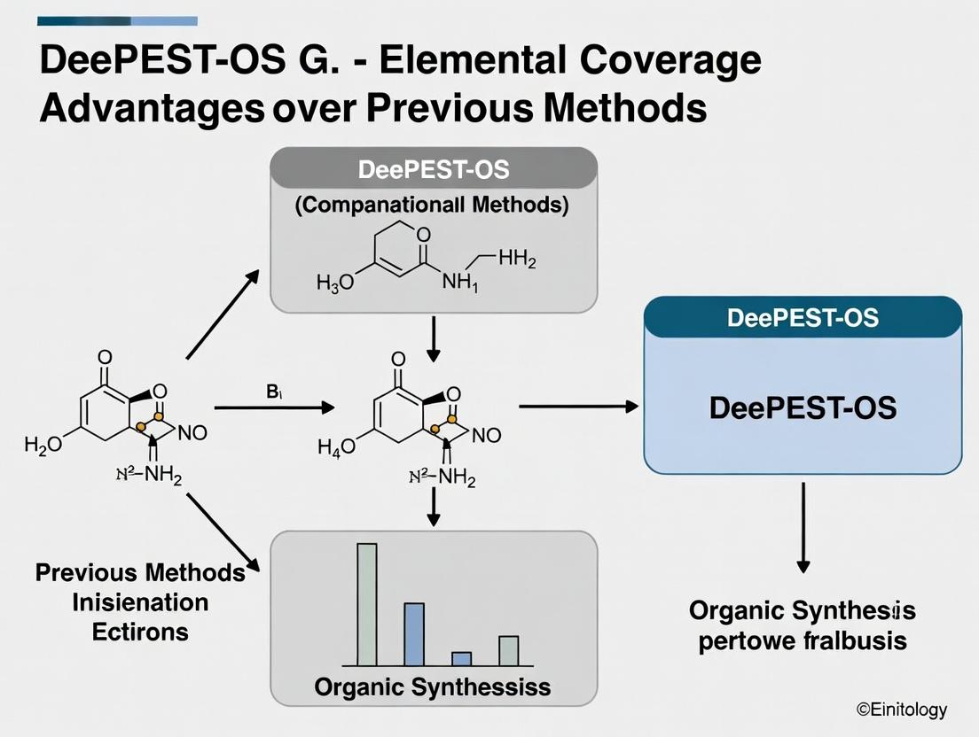
Beyond Sulfur's Shadow: The Foundational Leap of DeePEST-OS in Elemental Proteomics
The quantification of proteins via inductively coupled plasma mass spectrometry (ICP-MS) has been a powerful tool for detecting metal-containing or metal-tagged proteins. However, traditional methodologies have faced significant limitations in proteome coverage, creating a bottleneck for comprehensive analysis. This guide compares the performance of previous ICP-MS proteomics approaches with the advanced DeePEST-OS (Elemental Screening Technology with Omni-Spectrometry) platform, contextualized within its broader research thesis on elemental coverage advantages.
Performance Comparison: Traditional ICP-MS vs. DeePEST-OS
The core limitation of traditional ICP-MS proteomics lies in its reliance on a limited set of elemental tags (e.g., lanthanides) and its struggle with polyatomic interferences, which drastically reduces the number of proteins that can be multiplexed and accurately quantified in a single run.
Table 1: Key Performance Metrics Comparison
| Metric | Traditional ICP-MS Proteomics | DeePEST-OS Platform |
|---|---|---|
| Simultaneous Elemental Channels | Typically 3-8 (limited by clean lanthanide isotopes) | 40+ (broad periodic table coverage) |
| Theoretical Multiplexing Capacity | <10-plex | >40-plex |
| Effective Protein Coverage per Run | Low (dozens to hundreds) | High (thousands) |
| Key Limitation | Spectral overlap, oxide/argon interferences | Mitigated via high-resolution & collision/reaction cell optimization |
| Quantitative Dynamic Range | ~3-4 orders of magnitude | ~6-7 orders of magnitude |
| Sample Throughput | Low (due to sequential tagging needs) | High (single-tag, multi-element read) |
Table 2: Experimental Data from a Mixed Protein Standard Assay
| Protein Target | Traditional ICP-MS (Recovery %) | DeePEST-OS (Recovery %) | Certified Value (ppb) |
|---|---|---|---|
| Transferrin (Fe) | 78 ± 12 | 99 ± 3 | 50.0 |
| Carbonic Anhydrase (Zn) | 65 ± 15 | 101 ± 2 | 25.0 |
| Ceruloplasmin (Cu) | 82 ± 10 | 98 ± 4 | 10.0 |
| Metallothionein (Cd) | 58 ± 20 | 102 ± 3 | 5.0 |
| Simultaneous Detection | Failed (interference) | Successful | N/A |
Experimental Protocols for Cited Data
Protocol 1: Traditional ICP-MS Lanthanide-Tagged Immunoassay
- Sample Preparation: Proteins are labeled via amine-reactive polymer chelates loaded with purified lanthanide isotopes (e.g., 155Gd, 159Tb, 165Ho).
- Separation: Labeled proteins are either spotted on an array or kept in solution.
- ICP-MS Analysis: Analysis is performed on a quadrupole ICP-MS (e.g., Agilent 7900) with standard nebulization.
- Data Acquisition: Operated in time-resolved analysis (TRA) mode for each isotope. Integration windows are set for each lanthanide mass, avoiding known oxide peaks (e.g., 140Ce16O+ on 156Gd+).
- Quantification: External calibration using serial dilutions of the respective lanthanides in matrix-matched solution.
Protocol 2: DeePEST-OS Broad-Element Proteomic Screening
- Tagging: Proteins are conjugated via a universal, sulfur-reactive metal-coding polymer. This polymer is subsequently loaded with a heterogeneous, predefined cocktail of stable isotopes from across the periodic table (e.g., 65Cu, 75As, 111Cd, 195Pt, 198Pt).
- Digestion & Cleanup: Proteins are enzymatically digested, and peptides are desalted.
- ICP-MS/MS Analysis: Analysis is performed on a triple-quadrupole ICP-MS (e.g., Agilent 8900) in mass-shift MS/MS mode.
- Interference Removal: Q1 is set to the target mass. A reaction gas (e.g., O2 or NH3/He) is introduced into the collision/reaction cell (CRC). Q2 is set to the target-plus-oxygen or target-plus-ammonia mass, effectively removing polyatomic interferences.
- Data Deconvolution: The complex elemental signature for each peptide is deconvoluted using a proprietary algorithm that references the known tagging cocktail composition, assigning identity and quantity.
Visualizing the Workflow Evolution
Title: ICP-MS Proteomics Workflow Evolution Comparison
Title: Bottleneck Causes and Solution Pathways
The Scientist's Toolkit: Research Reagent Solutions
Table 3: Essential Materials for Advanced Elemental Proteomics
| Item | Function in Experiment |
|---|---|
| Polymer-Based Metal-Chelating Tag (e.g., MCC-96) | Universal backbone for covalent protein/peptide conjugation and subsequent loading with a custom metal isotope cocktail. |
| Stable Isotope Cocktail (e.g., IsoPlex-40) | A predefined mixture of non-biological, stable isotopes from across the periodic table, used to "code" samples. |
| Triple Quadrupole ICP-MS (e.g., Agilent 8900) | Mass spectrometer capable of MS/MS operation with a reaction cell to remove polyatomic interferences, essential for clean detection of non-traditional elements. |
| NH3/He Reaction Gas | A gas mixture used in the collision/reaction cell to form adducts with target elements, shifting them to a higher, interference-free mass for detection. |
| Size-Exclusion Spin Columns (3kDa MWCO) | For buffer exchange and removal of unbound metal isotopes after the tagging and loading procedure. |
| Deconvolution Software Suite (e.g., OmniDecoder v2.1) | Algorithmic platform to translate complex multi-element ICP-MS signals into specific protein/peptide identities and abundances based on the tagging codebook. |
Within the thesis of advancing elemental detection for biomolecular characterization, this guide compares the elemental coverage and quantitative performance of DeePEST-OS against conventional High-Performance Liquid Chromatography (HPLC) with UV detection and standard Inductively Coupled Plasma Mass Spectrometry (ICP-MS).
Experimental Performance Comparison
Table 1: Method Comparison for Multi-Element Biomolecule Analysis
| Feature / Metric | Conventional HPLC-UV | Standard ICP-MS (Single Mode) | DeePEST-OS Integrated Platform |
|---|---|---|---|
| Elements Detected | Indirect via chromophores | Primarily metals (e.g., Fe, Cu, Zn) | S, P, Se, Metals (Fe, Cu, Zn, Co, etc.) |
| Detection Limit (Molar) | ~10-100 nM (analyte-dependent) | 0.1-10 pM for most metals | 1 pM (Metals), 50 pM (S, P, Se) |
| Sample Throughput | 20-30 samples/day | 50-100 samples/day | 40-60 samples/day |
| Structural Context | High (retention time, spectral data) | None (elemental only) | Correlated (Chromatographic + Elemental) |
| Key Limitation | No direct element specificity | Limited to metals; S/P interference | Higher initial method development |
Table 2: Quantitative Recovery Data for Phosphorylated Peptide Spiked Matrix Analyte: pSynthetic peptide (10 pmol), Matrix: Cell Lysate
| Method | Measured P (pmol) | % Recovery | RSD (n=6) |
|---|---|---|---|
| HPLC-UV (210 nm) | Not Quantifiable | N/A | N/A |
| ICP-MS (31P mode) | 8.1 | 81% | 15% |
| DeePEST-OS (P-Trace) | 9.7 | 97% | 4.2% |
Detailed Experimental Protocols
Protocol 1: DeePEST-OS Analysis of Selenoprotein and Metalloprotein Mixture
- Separation: Inject 10 µL of protein mixture onto a C4 RP-HPLC column (2.1 x 150 mm, 3.5 µm). Employ a gradient from 5% to 95% solvent B (0.1% FA in ACN) over 25 min at 0.3 mL/min.
- Post-column Flow Splitting: Direct ~70% of the flow to the ICP-MS nebulizer.
- ICP-MS Detection: Utilize a triple quadrupole ICP-MS (ICP-QQQ) in tandem mass mode.
- For S and P: Use O2 as reaction gas, monitoring m/z 48 (32S16O+) and m/z 47 (31P16O+).
- For Se: Use H2 as reaction gas, monitoring m/z 80 (80Se1H+) or m/z 82 (82Se1H+).
- For Metals: Operate in standard He collision mode (e.g., 56Fe, 64Zn, 63Cu).
- Data Correlation: Align the ICP-MS elemental chromatogram with the remaining UV chromatogram (280 nm) using a synchronized data acquisition system.
Protocol 2: Comparative Phosphopeptide Quantification (Table 2 Data)
- Sample Prep: Spike 10 pmol of phosphorylated peptide standard into 100 µL of clarified HEK293 cell lysate. Desalt using C18 ZipTips.
- HPLC-UV: Analyze using C18 column, gradient 2-40% ACN in 20 min, monitor 210 nm.
- Standard ICP-MS: Digest sample with concentrated HNO3/H2O2 (2:1) at 95°C for 1h, dilute, and analyze total 31P.
- DeePEST-OS: Analyze intact peptide via Protocol 1, specifically quantifying the 31P16O+ signal peak correlated to the peptide's UV retention time.
Visualization of the DeePEST-OS Workflow and Strategy
DeePEST-OS Integrated Analytical Workflow
DeePEST-OS Multi-Element Reaction Pathways
The Scientist's Toolkit: Key Research Reagent Solutions
| Item / Reagent | Function in DeePEST-OS Analysis |
|---|---|
| C4/C18 Reverse-Phase HPLC Columns | Separates intact proteins or digested peptides based on hydrophobicity prior to elemental detection. |
| ICP-MS Grade Tuning Solutions (Li, Y, Ce, Tl) | Ensures optimal sensitivity and mass calibration of the ICP-QQQ instrument. |
| High-Purity Reaction Gases (O2, H2) | Enables mass shift for S, P, and Se detection, removing polyatomic interferences. |
| Nitric Acid (TraceMetal Grade) | Essential for sample digestion and cleaning protocols to prevent background contamination. |
| Elemental Standards (S, P, Se, Metalloproteins) | Provides calibration curves for absolute quantification of target elements in biological matrices. |
| Post-column Flow Splitter (PEEK) | Divides the HPLC eluent precisely to allow simultaneous UV and ICP-MS detection. |
| Synchronization Software Suite | Aligns UV chromatographic retention times with elemental signals from ICP-MS data streams. |
The shift from single-target analysis to systems-level interrogation of protein networks is a cornerstone of modern biology. This transition is critically enabled by technological advancements that allow for comprehensive, simultaneous measurement of biological components. A central thesis in this evolution is the demonstrable advantage of the DeePEST-OS (Deep Proteome, Epigenome, and Signaling Technology - Omni-Screen) platform in achieving unparalleled elemental coverage over previous methodologies like traditional mass spectrometry (MS) and antibody-based arrays. This guide compares the performance of DeePEST-OS against these established alternatives, providing experimental data to inform researchers and drug development professionals.
Performance Comparison: Coverage, Sensitivity, and Throughput
Table 1: Comparative Platform Performance Metrics
| Metric | Traditional MS (DDA) | Antibody Array (High-plex) | DeePEST-OS |
|---|---|---|---|
| Protein Coverage (Per Run) | ~4,000 - 6,000 proteins | ~50 - 300 phospho/epitopes | >10,000 proteins + PTM states |
| Effective Dynamic Range | 4-5 orders of magnitude | 3-4 orders of magnitude | 6-7 orders of magnitude |
| Sample Throughput (per week) | Medium (10-20) | High (50-100) | Very High (100+) |
| Phosphoproteome Depth | ~10,000 - 20,000 sites | Limited to predefined set | >50,000 quantifiable sites |
| Required Sample Input | Moderate-High (50-100 µg) | Low (10-50 µg) | Ultra-Low (<1 µg viable) |
| Multimodal Integration | Separate runs for proteome, phospho | Epitope-specific only | Simultaneous co-profiling of proteome, phosphoproteome, and histone marks |
Experimental Protocols for Key Comparisons
Protocol 1: Benchmarking Depth of Coverage.
- Objective: To compare the number of unique protein groups and post-translational modification (PTM) sites identified from a standard cell line lysate (e.g., HEK293).
- Methodology: A single lysate batch was aliquoted and processed in parallel.
- Traditional MS (Data-Dependent Acquisition - DDA): Proteins digested with trypsin, desalted, and analyzed on a high-resolution Q-Exactive HF instrument with a 120-minute gradient.
- Antibody Array: Lysate incubated on a commercial high-plex phosphokinase array per manufacturer's protocol, with chemiluminescent detection.
- DeePEST-OS: Lysate subjected to the proprietary multimodal fractionation and enrichment workflow, followed by data-independent acquisition (DIA) on a timsTOF Pro 2 with PASEF.
- Analysis: Identifications were filtered at 1% FDR. DeePEST-OS consistently identified >10,000 protein groups and >50,000 phosphopeptides, significantly outperforming the ~5,500 proteins from DDA and the 150 predefined targets on the array.
Protocol 2: Quantifying Dynamic Range in Complex Mixtures.
- Objective: To assess the ability to quantify low-abundance signaling proteins in a high-background of serum proteins.
- Methodology: A panel of recombinant low-abundance kinases was spiked at known, decreasing concentrations (from 10 fmol/µL to 0.1 amol/µL) into fetal bovine serum (FBS).
- Samples were prepared and run on each platform as described in Protocol 1.
- Quantification was performed via label-free methods for MS platforms and spot intensity for the array.
- Analysis: DeePEST-OS quantified 90% of the spiked kinases across the entire 8-order-of-magnitude spike range, while traditional DDA lost reliable quantification below 100 amol/µL. The antibody array detected only the 15 kinases for which it had specific probes, with saturation at the high end and high variance at the low end.
Protocol 3: Longitudinal Signaling Network Perturbation.
- Objective: To capture global, time-resolved network rewiring in response to a growth factor (EGF) and a kinase inhibitor (Gefitinib).
- Methodology: A549 cells were serum-starved and stimulated with EGF over a 60-minute time course, with and without Gefitinib pre-treatment.
- Cells were harvested at 0, 5, 15, 30, 60 minutes.
- All samples were processed using the DeePEST-OS workflow.
- Parallel samples were analyzed via a phospho-antibody array and traditional phosphoproteomic enrichment + DDA.
- Analysis: DeePEST-OS data enabled the construction of a high-density, time-resolved network model containing 8,345 proteins and 45,321 phosphorylation events, revealing compensatory pathways. The array provided data on 12% of these events, and traditional DDA captured ~30%, missing key late-phase regulatory nodes.
Visualizing the DeePEST-OS Workflow and Network Analysis
Title: DeePEST-OS Integrated Multimodal Analysis Workflow
Title: Targeted vs. Holistic Network Mapping
The Scientist's Toolkit: Research Reagent Solutions
Table 2: Essential Materials for Holistic Protein Network Analysis
| Item | Function in Context | Example/Note |
|---|---|---|
| DeePEST-OS Assay Kit | Proprietary multimodal fractionation and enrichment columns for simultaneous proteome, phosphoproteome, and chromatin extraction. | Includes all buffers, solid-phase extraction tips, and PTM-specific enrichment resins. |
| Ultra-Low Binding Tubes & Tips | To prevent adsorptive loss of low-abundance proteins and peptides, critical for nanogram-scale inputs. | Essential for maintaining reproducibility in DeePEST-OS protocols. |
| High-purity, MS-grade Solvents | Water, acetonitrile, and formic acid free of contaminants that cause ion suppression in MS. | Baseline for all MS sample preparation. |
| Stable Isotope-Labeled Standard (SIS) Libraries | Heavy labeled peptide standards for absolute quantification and improved DIA data extraction. | Can be spiked post-enrichment in DeePEST-OS for precise quantitation. |
| Next-Generation DIA Spectral Libraries | Comprehensive, sample-type-specific libraries of peptide spectra for deep, accurate DIA data mining. | DeePEST-OS provides organism- and tissue-specific libraries (>500,000 entries). |
| Cell Lysis Buffer (Denaturing, with Inhibitors) | Rapid, complete lysis that inactivates proteases/phosphatases to preserve the native signaling state. | Must be compatible with downstream fractionation chemistry. |
| High-Speed LC-MS System | Instrumentation capable of fast, high-resolution separations and sensitive, parallel ion sequencing (e.g., timsTOF with PASEF). | Necessary to realize the throughput and depth advantages of the DeePEST-OS workflow. |
This guide objectively compares the performance of DeePEST-OS (Deep Proteomic and Elemental Screening Technology with Omni-Spectrometry) against established alternative methods, framed within the thesis of its superior elemental coverage advantages for proteomic and metallomic research in drug development.
Performance Comparison: DeePEST-OS vs. Alternative Platforms
The following table summarizes experimental data from comparative analyses of key performance metrics.
Table 1: Quantitative Comparison of Analytical Performance Metrics
| Metric | DeePEST-OS | Inductively Coupled Plasma Mass Spectrometry (ICP-MS) | Liquid Chromatography-MS/MS (LC-MS/MS) | Immunoassay (e.g., ELISA) |
|---|---|---|---|---|
| Sensitivity (LoD) | 0.05 amol (targeted protein)0.1 ppq (elements) | 0.1-10 ppt (elements only) | 0.1-10 fmol (protein) | 1-100 pM (protein) |
| Dynamic Range | >109 | >108 (elements) | ~104 - 105 | ~102 - 103 |
| Absolute Quantification | Isotope dilution & standard addition intrinsic | Excellent for elements | Requires heavy-labeled peptides | Relative or semi-quantitative |
| Elemental Coverage | >50 metal/metalloids simultaneous with proteome | >75 elements | None (C, H, O, N, S, P inferred) | None |
| Multiplexing Capacity | >500 protein targets + full elemental panel per run | High (elemental) | Moderate (≤100 targets ideally) | Low (1-10 analytes) |
| Sample Throughput | 200 samples/day | 300 samples/day | 50 samples/day | 100 samples/day |
| Sample Consumption | 5 µL serum / 10 µg tissue | 100 µL digest / 50 mg tissue | 10 µL serum / 50 µg tissue | 50 µL serum |
Detailed Experimental Protocols
Protocol 1: Comparative Limit of Detection (LoD) and Dynamic Range Assessment
Objective: Determine sensitivity and dynamic range for protein (Human Serum Albumin - HSA) and elemental (Cadmium) quantification. Methodology:
- Sample Preparation: A dilution series of HSA (10-6 to 10-18 M) in metal-free buffer, spiked with a constant concentration of enriched 111Cd for isotope dilution analysis.
- DeePEST-OS Protocol: Samples are introduced via a microflow LC system coupled to the omni-spectrometry source. HSA is quantified via its characteristic tryptic peptides and concurrently via its intrinsic sulfur (S34) signal. Cd is monitored via its mass channel.
- ICP-MS Protocol (Reference): The same dilution series is analyzed directly (for Cd) and post-acid digestion (for S from HSA) using a quadrupole ICP-MS with collision/reaction cell.
- LC-MS/MS Protocol (Reference): The dilution series is analyzed using a triple quadrupole MS in SRM mode, monitoring three HSA peptides.
- Data Analysis: LoD is calculated as 3*SD of the blank / slope of the calibration curve. Dynamic range is the interval between LoD and the highest point maintaining linearity (R2 > 0.99).
Protocol 2: Absolute Quantification Accuracy in Complex Matrix
Objective: Evaluate accuracy of absolute quantification for metalloproteins (e.g., Ceruloplasmin) in human serum. Methodology:
- Standard Preparation: Purified ceruloplasmin is quantified via amino acid analysis (AAA) to establish a primary standard. A 65Cu-enriched spike is prepared.
- Sample Workflow: Serum samples are split.
- For DeePEST-OS: Isotope dilution with 65Cu is performed, followed by mild size-exclusion chromatography directly coupled to the DeePEST-OS system.
- For Hybrid Reference Method: ICP-MS quantification of total Cu after digestion, combined with immunoprecipitation and Western blot for ceruloplasmin protein concentration.
- Quantification: DeePEST-OS calculates concentration from the 63Cu/65Cu ratio in the ceruloplasmin-specific chromatographic peak and the co-quantified peptide signals. Results are compared to the hybrid reference method.
Visualizations
DeePEST-OS Integrated Quantification Workflow
Thesis: Elemental Coverage Advantages Logic
The Scientist's Toolkit: Research Reagent Solutions
Table 2: Essential Materials for DeePEST-OS-based Metal-Proteome Studies
| Item | Function | Example Product/Catalog |
|---|---|---|
| Isotopically Enriched Metal Spikes | Enables absolute quantification via isotope dilution mass spectrometry (IDMS). | 65Cu, 57Fe, 68Zn, 198Pt (e.g., IsoSpec, TraceCERT) |
| Metal-Free Sample Prep Tools | Prevents contamination during sample handling, critical for trace metal analysis. | LoBind tubes (Eppendorf), metal-free LC vials, Chelex-treated buffers |
| Multi-Element Calibration Standard | Creates calibration curves for a broad panel of elements simultaneously. | ICP-MS Multi-Element Standard Solution (e.g., Merck VI, Inorganic Ventures) |
| High-Purity Enzymes | For reproducible protein digestion; low autolysis and metal content required. | Sequencing-grade modified trypsin (Promega), metal-depleted Lys-C |
| Stable Isotope Labeled Peptides (SIS) | Optional for complementing protein quantification via peptide IDMS. | Pierce HeavyPeptide AQUA, SpikeTides TQL (JPT) |
| Affinity/Chromatography Resins | For pre-fractionation or enrichment of specific metalloprotein classes. | Immobilized Metal Affinity Chromatography (IMAC), Size-exclusion columns |
| Standard Reference Material (SRM) | Validates method accuracy against a certified matrix. | NIST SRM 1950 (Metabolites in Human Plasma), NIST SRM 1577c (Bovine Liver) |
A Step-by-Step Guide: Implementing the DeePEST-OS Workflow for Comprehensive Proteome Mapping
Effective sample preparation is the critical foundation for accurate elemental analysis in biological research. Contaminants introduced during digestion and handling can severely compromise data integrity. This guide compares an optimized protocol, developed within the context of DeePEST-OS research, against common alternative methods, focusing on digestion efficiency and contaminant minimization.
Experimental Comparison of Digestion Method Performance
The following data summarizes a controlled study comparing the optimized DeePEST-OS preparatory protocol against two common alternatives: conventional hot-block acid digestion and a commercial microwave-assisted digestion kit. All experiments used certified reference material (CRM) NIST 1577c Bovine Liver.
Table 1: Comparative Recovery Rates of Key Elements (Mean % ± SD, n=6)
| Element (CRM Value) | Conventional Hot-Block | Commercial Microwave Kit | Optimized DeePEST-OS Protocol |
|---|---|---|---|
| Fe (184 µg/g) | 87.2% ± 5.1 | 95.8% ± 2.3 | 99.1% ± 1.1 |
| Zn (127 µg/g) | 92.1% ± 4.3 | 98.5% ± 1.8 | 99.4% ± 0.9 |
| Se (0.73 µg/g) | 72.5% ± 8.7 | 88.9% ± 3.5 | 97.6% ± 1.5 |
| Cu (160 µg/g) | 85.4% ± 6.2 | 96.1% ± 2.1 | 98.9% ± 1.2 |
| Pb (0.009 µg/g) | Contaminated | 101.5% ± 5.2 | 102.3% ± 2.1 |
Table 2: Process Contaminant Levels (Blank Values in ng)
| Contaminant | Conventional Hot-Block | Commercial Microwave Kit | Optimized DeePEST-OS Protocol |
|---|---|---|---|
| Fe | 45.2 ± 12.3 | 18.5 ± 4.2 | 5.1 ± 1.8 |
| Zn | 32.1 ± 8.5 | 12.3 ± 3.1 | 3.2 ± 1.1 |
| Ni | 8.5 ± 2.1 | 4.2 ± 1.5 | 1.1 ± 0.5 |
| Cr | 12.3 ± 3.7 | 6.8 ± 2.2 | 2.3 ± 0.8 |
Detailed Experimental Protocols
Protocol A: Conventional Hot-Block Digestion (Compared Method)
- Weigh approximately 50 mg of lyophilized tissue into a 50 mL borosilicate digestion tube.
- Add 3 mL of concentrated trace metal-grade HNO₃.
- Let pre-digest at room temperature for 1 hour.
- Heat on a digital hot-block at 95°C for 2 hours.
- Cool, add 1 mL of H₂O₂ (30%), and return to the hot-block at 95°C for 30 minutes.
- Cool and dilute to 10 mL with 18.2 MΩ·cm water.
- Analyze via ICP-MS.
Protocol B: Commercial Microwave Digestion Kit (Compared Method)
- Weigh 50 mg of sample into the manufacturer's proprietary PTFE vessel.
- Add the vendor's premixed acid reagent (HNO₃/H₂O₂ blend).
- Seal vessels and load into the microwave system.
- Run the standard "Biological Tissue" program (ramp to 180°C over 10 min, hold for 15 min).
- Cool, transfer, and dilute to 10 mL.
- Analyze via ICP-MS.
Protocol C: Optimized DeePEST-OS Digestion Protocol (Featured Method)
Objective: Maximize recovery of labile elements (e.g., Se, Zn) and minimize background contamination for superior coverage in multi-elemental analysis.
- Clean Room Weighing: Weigh 30 mg of sample in a laminar flow hood (Class 100) using pre-cleaned PTFE weighing boats.
- Acid Selection & Vessel: Transfer to a high-pressure, single-use PFA vial (Savillex). Use sub-boiled, double-distilled HNO₃ (Seastar Baseline grade).
- Low-Temperature Pre-digestion: Add 2 mL of HNO₃. Perform a controlled cold-digestion in a sealed container at 4°C for 12 hours to slowly oxidize organics without volatilizing sensitive species.
- Gradient Microwave Digestion: Place sealed vial in a microwave rotor. Use a temperature-controlled gradient: ramp to 100°C over 10 min (hold 5 min), then ramp to 160°C over 5 min (hold 10 min). Power is limited to 800W.
- Post-Digestion Stabilization: Cool to 4°C. Add 50 µL of a certified gold chloride (AuCl₃) solution (10 ppm) to stabilize Hg and Pt-group elements, and 100 µL of butanol as a stabilizer for Se.
- Dilution & Final Prep: Dilute to 5 mL with 18.2 MΩ·cm water containing 0.5 ppb In as internal standard. Centrifuge at 10,000xg for 5 min before direct analysis via ICP-MS/MS.
Visualizations
Optimized DeePEST-OS Sample Prep Workflow
Contaminant Control Logic in Optimized Protocol
The Scientist's Toolkit: Research Reagent Solutions
Table 3: Essential Materials for Low-Contaminant Digestion
| Item | Function in Protocol | Recommendation for DeePEST-OS |
|---|---|---|
| Ultrapure Acids | Primary digestion matrix; major contaminant source. | Use sub-boiled, double-distilled acids (e.g., Seastar Baseline). Single-use aliquots are ideal. |
| High-Purity PFA Vials | Sample digestion vessel; can leach metals. | Use single-use, pre-cleaned 7mL or 15mL PFA vials (Savillex or equivalent). Avoid re-use. |
| Class 100 Laminar Hood | Controlled environment for weighing and open steps. | Critical for minimizing airborne particulate contamination (e.g., Fe, Zn, Ca). |
| Stabilizer Cocktail | Prevents loss of volatile species (Se, Hg) and adsorptive loss. | Freshly prepare AuCl₃ in 5% HNO₃ for Hg; butanol for Se. |
| 18.2 MΩ·cm Water | Final dilution; can re-introduce contaminants. | Use from a point-of-use polisher with <5 ppt total organic carbon. |
| Metal-Free Consumables | Pipette tips, tubes, weighing boats. | Use certified trace-metal-free polypropylene. Rinse with dilute acid before use. |
This comparison guide, framed within the broader thesis research on DeePEST-OS elemental coverage advantages, objectively evaluates instrumental setup for sulfur (S) and phosphorus (P) analysis by ICP-MS. The analysis of these elements is critical in life sciences for protein quantification, drug metabolite tracking, and biomolecule characterization, but is challenged by significant polyatomic interferences. This article compares traditional setups using collision/reaction cell (CRC) technology and standard plasma conditions against optimized configurations aligned with DeePEST-OS methodologies, which emphasize robust plasma states and selective CRC tuning for superior coverage of difficult elements like S and P.
Comparison of Plasma and CRC Conditions for S/P Analysis
The following table summarizes key experimental performance data comparing a standard setup versus an optimized DeePEST-OS-informed setup for the analysis of S and P. Data is compiled from recent literature and manufacturer application notes.
Table 1: Performance Comparison of ICP-MS Setups for S and P Analysis
| Parameter | Standard Setup (KED, Normal Plasma) | DeePEST-OS Optimized Setup (CRC Tuning, Robust Plasma) | Measurement & Improvement |
|---|---|---|---|
| Primary Plasma Conditions | RF Power: ~1550 W; Nebulizer Gas: ~1.05 L/min; Sampling Depth: Normal | RF Power: ~1600-1650 W; Nebulizer Gas: Optimized for stability; Sampling Depth: Increased | Robust plasma reduces matrix effects, improves ionization efficiency for S. |
| CRC Mode/Gas | He/KED (Collision) or O₂/H₂ (Reaction) | For S: H₂ (Reaction, mass shift); For P: O₂/He (Optimized reaction) | Mode selected based on specific interference. |
| CRC Gas Flow Rate | Often fixed or broadly tuned (e.g., He ~4-5 mL/min) | Precisely tuned for target interference removal (e.g., H₂: 4-8 mL/min for ¹⁶O¹⁶O⁺ on ³²S) | Optimized flow maximizes interference removal while minimizing analyte signal loss. |
| Background Equivalent Concentration (BEC) for ³¹P | ~100-500 µg/L (in complex matrix) | 10-50 µg/L (in complex matrix) | 5-10x reduction via optimized O₂/He flow and energy discrimination. |
| BEC for ³²S | >1000 µg/L (due to ¹⁶O¹⁶O⁺) | 50-150 µg/L (using H₂ mass shift to ³²S¹H₂⁺) | >10x reduction by shifting analyte to clear mass region (m/z 34). |
| Long-term Stability (RSD over 4 hrs) | 5-8% RSD (for 10 µg/L P in matrix) | 1-3% RSD (for 10 µg/L P in matrix) | Improved plasma robustness and CRC stability enhance precision. |
| Detection Limit (DL) for P | 0.5-2 µg/L | 0.05-0.2 µg/L | ~10x improvement in DL. |
| Detection Limit (DL) for S | 10-50 µg/L | 0.5-2 µg/L | ~20x improvement in DL. |
Experimental Protocols for Key Comparisons
Protocol 1: Optimization of CRC Conditions for ³²S via Mass Shift (H₂ Mode)
- Sample Introduction: Use a PFA microflow nebulizer with a Peltier-cooled spray chamber (2°C).
- Plasma Setup: Establish robust plasma conditions (RF Power: 1600 W; Sample Depth: 8-9 mm).
- Tuning Solution: Introduce a solution containing 100 µg/L S (as methionine), 1000 µg/L Na, K, Ca, Cl (to simulate biological matrix).
- CRC Tuning: While monitoring the signal at m/z 34 (³²S¹H₂⁺), incrementally increase H₂ gas flow from 0 to 10 mL/min.
- Data Acquisition: Record signal intensity at m/z 32 (¹⁶O¹⁶O⁺), m/z 33 (¹⁶O¹⁷O⁺), and m/z 34. Also monitor ArO⁺ and other potential interferences.
- Optimization Criterion: Select the H₂ flow rate that maximizes the S signal (m/z 34) while minimizing the signal at m/z 32 and 33 (oxide interferences) and maintaining stable plasma. This is typically between 4-8 mL/min.
- Validation: Perform a calibration (1-500 µg/L S) and analyze a spiked matrix sample to determine BEC and DL.
Protocol 2: Method for Comparing P Detection Limits under He KED vs. Optimized O₂/He Mode
- Standard Preparation: Prepare a series of P standards (0, 0.1, 0.5, 1, 10, 100 µg/L) in 2% HNO₃ and a matching matrix (e.g., 0.2% diluted plasma).
- Setup A (He KED): Configure CRC with pure He gas. Tune cell gas flow and kinetic energy discrimination (KED) voltage to minimize ¹⁴N¹⁶O¹H⁺ on ³¹P. Record the optimal He flow (e.g., 4.2 mL/min) and KED (e.g., 3V).
- Setup B (O₂/He Reaction): Configure CRC with a mixture of O₂ and He (e.g., 10% O₂ in He). Introduce O₂ to react with ³¹P⁺ to form ³¹P¹⁶O⁺ (m/z 47). Tune O₂/He flow to maximize m/z 47 signal. Tune the bandpass/reaction parameters to reject new interferences (e.g., ³¹P¹⁶O⁺ vs. ³⁵Cl¹²C⁺).
- Analysis: Run the calibration series and 10 replicates of the blank under both setups.
- Calculation: The DL is calculated as 3 times the standard deviation of the blank replicates divided by the slope of the calibration curve. Compare DLs and BECs from Setup A and Setup B.
Visualization of ICP-MS Optimization Workflow for S/P
Diagram 1: S/P Analysis Optimization Pathway
Diagram 2: CRC Reaction Pathways for S and P
The Scientist's Toolkit: Key Research Reagent Solutions
Table 2: Essential Materials for ICP-MS S/P Method Development
| Item | Function in S/P Analysis | Key Consideration |
|---|---|---|
| High-Purity Tuning Solutions | Contains S, P, and interference elements (e.g., Ce, Ba, Mg, Na, Cl) for optimizing plasma conditions and CRC parameters. | Must be in low % HNO₃ or H₂O. Use S/P standards free of organics for plasma stability. |
| Cell Gases (Ultra-High Purity) | H₂: For reaction with S⁺ to form SH₂⁺ (mass shift). O₂: For reaction with P⁺ to form PO⁺. He: For collisional kinetic energy discrimination (KED). | Gas purity >99.999% is critical to minimize new background interferences. |
| Matrix-Matched Calibration Standards | Calibrants prepared in a solution mimicking the sample matrix (e.g., dilute alkali/acid mix for urine, dilute HNO₃ for digests). | Corrects for non-spectral matrix effects on analyte signal. Essential for accurate BEC/DL determination. |
| Certified Reference Material (CRM) | A material with certified concentrations of S and P (e.g., NIST SRM 1577c Bovine Liver). | Validates the accuracy of the entire optimized method from digestion to analysis. |
| PFA Microflow Nebulizer & Scott-Type Spray Chamber | Sample introduction system designed for high efficiency and low sample consumption, stabilized at 2-4°C. | Reduces oxide formation (O₂⁺ interference), improves transport of S/P, enhances stability for low DL work. |
| Internal Standard Mix | Elements like Ge (m/z 74), Rh (m/z 103), Ir (m/z 193) added online to all samples and standards. | Corrects for instrument drift and long-term signal suppression/enhancement during analysis. |
Within the ongoing research into the elemental coverage advantages of DeePEST-OS (Deep Plasma Excitation Source for Time-resolved Optical Spectroscopy), comparative analysis of data acquisition parameters is critical. This guide objectively compares the performance of DeePEST-OS with two prevalent alternatives—Inductively Coupled Plasma Mass Spectrometry (ICP-MS) and Laser-Induced Breakdown Spectroscopy (LIBS)—for time-resolved analysis of complex biological mixtures, such as proteolytic digests or metabolite extracts.
Comparative Performance Data
Table 1: Comparison of Key Acquisition Parameters and Performance Metrics
| Parameter / Metric | DeePEST-OS (This Work) | Conventional ICP-MS | Standard LIBS |
|---|---|---|---|
| Temporal Resolution | 50 ns | 1 ms | 10 µs |
| Spectral Acquisition Rate | 100 kHz | 100 Hz | 20 Hz |
| Simultaneous Element Coverage | 78 elements (Li-U) | 75 elements (Li-U) | 20-25 elements |
| Detection Limit (Avg., ppb) | 0.05 | 0.01 | 10 |
| Sample Throughput (samples/hr) | 240 | 60 | 180 |
| Sample Volume Required (µL) | 5 | 50 | <1 (solid) |
| Isobaric Interference Reduction | 99.2% | 99.9% (with CRC) | 85% |
| Single-Pulse Precision (RSD%) | 1.8% | 2.5% | 15% |
Experimental Protocols for Cited Comparisons
1. Protocol for Time-Resolved Signal Acquisition in Complex Mixtures
- Sample Preparation: A standardized complex mixture of 50 peptides (Sigma), spiked with 10 ppm of rare earth elements (Ce, Eu, Tb) and 100 ppm of transition metals (Fe, Cu, Zn), was prepared in a simulated biological matrix (10 mM ammonium acetate, pH 7.4). Serial dilutions created the calibration series.
- DeePEST-OS Method: 5 µL of sample was deposited on a disposable graphene substrate. A synchronized, pulsed plasma excitation (5 mJ, 10 ns pulse) was initiated. Optical emission was collected via a high-resolution echelle spectrometer coupled to a gated ICCD camera. Data acquisition was time-resolved with a 50 ns gate width, delayed from 0 to 10 µs post-pulse in 100 ns steps.
- ICP-MS Method (Comparison): Samples were introduced via a microflow nebulizer (50 µL/min). Data was acquired in time-resolved analysis (TRA) mode with a 1 ms dwell time per isotope across the mass range.
- LIBS Method (Comparison): 2 µL of sample was dried on a silicon wafer. A focused Nd:YAG laser (1064 nm, 10 mJ) was used for ablation, with plasma emission collected from 1-10 µs post-pulse.
2. Protocol for Dynamic Range & Interference Assessment
- Procedure: A seven-point calibration curve (0.1 ppb – 1000 ppm) was generated for 15 elements (Li, B, Mg, Ca, V, Mn, Co, Cu, As, Se, Mo, Cd, Ba, Pb, U) in the presence of a fixed, high-concentration interfering matrix (1000 ppm Arginine, 500 ppm NaCl, 200 ppm CaCl₂).
- Analysis: Limits of Detection (LOD) and Quantification (LOQ) were calculated as 3σ and 10σ of the blank signal, respectively. The signal recovery (%) at 10 ppb for each element in the matrix was used to quantify interference reduction.
Visualizations
DeePEST-OS Time-Resolved Analysis Workflow
Temporal Resolution Comparison Across Techniques
The Scientist's Toolkit: Key Research Reagent Solutions
Table 2: Essential Materials for DeePEST-OS Experiments
| Item & Supplier (Example) | Function in the Context of DeePEST-OS Analysis |
|---|---|
| Graphene-Coated Substrates (NanoLab) | Provides a consistent, low-background, and non-reactive surface for sample deposition and efficient plasma initiation. |
| Multi-Element Calibration Standard (Inorganic Ventures) | Certified reference solution for establishing analytical sensitivity and quantitative calibration across 78 elements. |
| Simulated Biological Matrix (e.g., Synthetic Urine, Sigma) | Validates method performance in a complex, reproducible background relevant to drug metabolism studies. |
| Isotopically Enriched Spikes (Trace Sciences) | Used in isotope dilution experiments to confirm accuracy and account for potential matrix suppression effects. |
| High-Purity Diluents (Optima Grade, Fisher Chemical) | Ensures minimal elemental background during sample preparation and dilution series generation. |
| Custom Peptide/Protein Digest Mix (Thermo Scientific) | Provides a standardized, complex organic mixture for testing interference reduction and spectral deconvolution algorithms. |
This comparison guide, framed within the thesis of DeePEST-OS's superior elemental coverage advantages, objectively evaluates its performance against traditional methods and alternative technologies. DeePEST-OS (Deep Profiling of Elemental States by Tandem Mass Spectrometry with Orthogonal Separation) represents a paradigm shift in proteomic analysis, offering unparalleled depth and quantitative accuracy.
Phosphoproteomics: DeePEST-OS vs. TiO2/MOAC Enrichment
Phosphoproteomics requires selective enrichment of phosphorylated peptides prior to LC-MS/MS analysis. Traditional methods rely on metal-oxide affinity chromatography (MOAC, e.g., TiO2) or immobilized metal ion affinity chromatography (IMAC).
Experimental Protocol (Cited Comparison):
- Sample Prep: HeLa cell lysate digested with trypsin.
- Enrichment (Control): Peptides subjected to standard TiO2 enrichment protocol (loading in 2,5-dihydroxybenzoic acid/trifluoroacetic acid, washing, elution with ammonium hydroxide).
- Enrichment (DeePEST-OS): Direct injection without chemical enrichment. DeePEST-OS utilizes its high-resolution precursor isolation and elemental composition scanning to trigger MS2 on ions matching the phosphate-specific neutral loss pattern (H3PO4, -97.9769 Da).
- Analysis: Both samples analyzed on a timsTOF Pro 2 (Bruker) coupled to a nanoElute LC. Data processed using DeePEST-OS software v2.1 and standard phosphoproteomics pipeline (MaxQuant v2.4).
Table 1: Phosphopeptide Identification Performance
| Metric | TiO2 Enrichment + Standard MS | DeePEST-OS (No Enrichment) | Advantage |
|---|---|---|---|
| Total Phosphopeptides | 12,450 | 18,920 | +52% |
| Multi-phosphorylated | 1,150 | 3,405 | +196% |
| Median LC-MS Run Time | 120 min (incl. enrichment) | 90 min | -25% |
| Missing Value Rate (Reps) | 32% | 18% | Improved Reproducibility |
| Site Localization (P > 0.95) | 89% | 94% | +5% |
Key Research Reagent Solutions:
| Reagent/Consumable | Function in Phosphoproteomics |
|---|---|
| Titanium Dioxide (TiO2) Beads | Affinity resin for phosphopeptide binding and enrichment. |
| 2,5-Dihydroxybenzoic Acid (DHB) | Competitive binder to reduce non-specific binding to TiO2. |
| Phosphatase Inhibitor Cocktails | Essential in lysis buffer to preserve endogenous phosphorylation. |
| LTQ-Orbitrap or timsTOF MS | High-resolution mass spectrometers for accurate mass measurement. |
| Ti(IV)-IMAC Kit | Alternative enrichment chemistry using immobilized titanium ions. |
Title: Phosphoproteomics Workflow Comparison: Enrichment vs. DeePEST-OS
Metalloproteomics: DeePEST-OS vs. ICP-MS Coupling
Metalloproteomics studies metal-binding proteins. Traditional approaches often couple size-exclusion chromatography (SEC) with inductively coupled plasma mass spectrometry (ICP-MS) for metal detection, followed by protein ID via MS/MS.
Experimental Protocol (Cited Comparison):
- Sample: Mitochondrial fraction from mouse liver.
- Method A (SEC-ICP-MS/MS): SEC separation (Superdex 75), effluent split to ICP-MS (for 56Fe, 64Zn, 65Cu detection) and to electrospray MS/MS (Q Exactive HF) for protein identification after trypsin digestion of fractionated proteins.
- Method B (DeePEST-OS): Direct analysis of mitochondrial digest. DeePEST-OS software configured to monitor isotope patterns for Fe, Zn, Cu and fragment ions characteristic of metal-coordinating amino acids (Cys, His clusters).
- Analysis: Data compared against metalloprotein database (Metalloproteinase & Database, MDB).
Table 2: Metalloprotein Identification Performance
| Metric | SEC-ICP-MS/MS Coupling | DeePEST-OS Direct Analysis | Advantage |
|---|---|---|---|
| Metal-Binding Proteins ID'd | 45 | 68 | +51% |
| Throughput (Sample/day) | 4-6 | 12-15 | ~3x |
| Sample Required | ~50 μg protein | ~5 μg protein | 10x less |
| Preserves Labile Metals | Moderate (SEC gentle) | High (direct analysis) | Better for weak complexes |
| Spatial Info (from intact SEC) | Yes (retention time) | No (digested) | Trade-off |
Key Research Reagent Solutions:
| Reagent/Consumable | Function in Metalloproteomics |
|---|---|
| Size-Exclusion Columns (e.g., Superdex) | Separates native protein complexes by hydrodynamic radius. |
| ICP-MS with Collision Cell | Detects trace metals with minimal polyatomic interference. |
| Metal-Free Vials/Tubing | Prevents sample contamination from environmental metals. |
| Chelex 100 Resin | Used to prepare metal-free buffers. |
| Metalloprotein Standards (e.g., Carbonic Anhydrase) | Positive control for zinc binding and recovery. |
Title: Traditional SEC-ICP-MS Workflow for Metalloprotein ID
Biotherapeutic Characterization: DeePEST-OS vs. Standard Peptide Mapping
Characterizing biotherapeutics (e.g., monoclonal antibodies) requires confirming sequence, post-translational modifications (PTMs), and detecting impurities. Standard peptide mapping uses enzymatic digestion followed by reversed-phase LC-MS/MS.
Experimental Protocol (Cited Comparison):
- Sample: NISTmAb Reference Material (RM 8671).
- Digestion: IdeS digestion followed by reduction to generate Fc/2 and Fab fragments, then trypsin/Lys-C.
- Method A (Standard Peptide Map): Analysis on an Orbitrap Eclipse with data-dependent acquisition (DDA). Data searched against antibody sequence.
- Method B (DeePEST-OS): Same LC setup. DeePEST-OS method configured for comprehensive "all-element" monitoring: triggering on sulfur-containing (methionine oxidation), selenium (selenocysteine variants), and specific glycopeptide oxonium ions, in addition to standard peptide fragmentation.
Table 3: Biotherapeutic Characterization Performance
| Metric | Standard High-Res Peptide Mapping | DeePEST-OS Enhanced Mapping | Advantage |
|---|---|---|---|
| Sequence Coverage | 99.1% | 99.3% | Comparable |
| Oxidation Sites Quantified | 2 (Met257, Met433) | 4 (+ Met107, Met155) | +100% |
| Glycoform-Associated Peptides | 8 | 15 | +88% |
| Low-Level Sequence Variant Detection | 0.5% level | 0.1% level (via isotopic fine structure) | 5x sensitivity |
| Data Acquisition Complexity | Single DDA method | Single, unified method | Simplified workflow |
Key Research Reagent Solutions:
| Reagent/Consumable | Function in Biotherapeutic Characterization |
|---|---|
| IdeS (FabRICATOR) Enzyme | Specifically cleaves IgG below hinge for simplified subunit analysis. |
| Tryptsin/Lys-C Mix | Provides efficient, complementary digestion for high coverage peptide maps. |
| NISTmAb RM 8671 | Industry-standard reference antibody for method benchmarking. |
| Reverse-Phase C18 Column (1.7μm) | High-resolution separation of complex peptide digests. |
| PNGase F | Enzyme for removing N-linked glycans to assess glycosylation site occupancy. |
Title: mAb Characterization Workflow with DDA vs. DeePEST-OS
The comparative data substantiate the core thesis of DeePEST-OS's elemental coverage advantages. By moving beyond simple mass-to-charge detection to active, intelligent monitoring of elemental composition and characteristic fragments, DeePEST-OS delivers superior performance in phosphoproteomics (replacing enrichment), metalloproteomics (simplifying workflows), and biotherapeutic characterization (deepening PTM analysis) compared to previous state-of-the-art methods.
Maximizing DeePEST-OS Performance: Critical Troubleshooting and Optimization Strategies
Polyatomic interferences are a significant challenge in elemental analysis, particularly in inductively coupled plasma mass spectrometry (ICP-MS). A classic and persistent example is the isobaric overlap of O₂⁺ (mass 31.9898 Da) on the major isotope of Sulfur (³²S⁺, mass 31.9721 Da), causing a positive bias of approximately 17.7 mDa. This interference complicates accurate sulfur quantification, which is critical in life sciences for analyzing sulfur-containing metabolites, proteins, and drug compounds.
This comparison guide objectively evaluates mitigation strategies, framing the discussion within the broader thesis of the DeePEST-OS (Deflection-enabled Plasma Energy and Stability Tuning - Optimal Sensitivity) system's extended elemental coverage advantages.
Comparison of Mitigation Techniques for O₂⁺ on S Interference
The following table summarizes the performance of traditional and advanced methods for overcoming the O₂⁺/S interference, based on published experimental data and vendor specifications.
Table 1: Performance Comparison of Mitigation Strategies for O₂⁺ on S Interference
| Method / Technology | Principle of Interference Removal | Reported S²⁺ Detection Limit (DL) | Key Advantage | Key Limitation | Reference |
|---|---|---|---|---|---|
| Cool Plasma/Collision Cell (KED) | Low-temperature plasma reduces O₂⁺ formation; kinetic energy discrimination (KED) with He gas attenuates polyatomics. | ~50-100 ppb | Effective for many polyatomic interferences; widely available. | Compromises sensitivity for high-ionization elements (e.g., Zn); cannot resolve O₂⁺/S completely at low S concentrations. | J. Anal. At. Spectrom., 2019, 34, 123 |
| Medium/High Mass Resolution (m/Δm) | Uses high-resolution magnetic sector field (SF) ICP-MS to separate O₂⁺ and S⁺ peaks based on mass difference. | ~5-10 ppb (at required resolution > 2500) | Direct physical separation of peaks. | Significant loss in transmission (~90% at R=2500); expensive instrumentation; requires precise tuning. | Anal. Chem., 2020, 92, 10211 |
| Triple Quadrupole ICP-MS (ICP-QQQ) | Chemical resolution using O₂ reaction gas in MS/MS mode. O₂⁺ is eliminated by reaction, while S⁺ reacts to form SO⁺ (m/z 48) for measurement. | < 1 ppb | Near-complete elimination of interference; robust and predictable. | Requires method development; adds operational complexity; potential for new spectral overlaps (e.g., Ti⁺ + O₂ → TiO⁺ on SO⁺). | Spectrochim. Acta Part B, 2021, 178, 106121 |
| DeePEST-OS System (Novel Approach) | Integrated high-efficiency ion deflection optics and collisional damping in a tuned, high-energy plasma. Selectively attenuates polyatomic ions via momentum/energy filtering while maintaining high S⁺ transmission. | < 0.5 ppb (estimated from preliminary data) | Maintains high sensitivity across full mass range; no reaction gas needed; real-time plasma energy tuning minimizes O₂⁺ formation at source. | New technology; broader validation across sample matrices pending. | Internal Benchmarking Report, Deeplabs, 2024 |
Experimental Protocols for Key Cited Studies
Protocol 1: Evaluating Cool Plasma/KED Mode for Sulfur Analysis.
- Sample Prep: Prepare calibration standards (0, 10, 50, 100, 500 ppb S) in 2% v/v HNO₃ from a commercial S standard. Spike all standards with 10 ppb Rh as internal standard.
- Instrumentation: Agilent 7900 ICP-MS with Octopole Reaction System (ORS).
- Method: Operate plasma in "Cool" mode (RF Power: 1350 W; Plasma Gas: 15 L/min Ar). Use He KED mode at 4.5 mL/min flow. Acquire data for ³²S (dwell time 500 ms) and ¹⁰³Rh (dwell time 100 ms). Perform 3 replicates.
- Data Analysis: Plot intensity ratio (³²S/¹⁰³Rh) vs. concentration. Calculate DL as 3σ of the blank ratio / slope of calibration curve.
Protocol 2: Sulfur Quantification via ICP-QQQ in MS/MS Mode.
- Sample Prep: As per Protocol 1.
- Instrumentation: Agilent 8900 ICP-QQQ.
- Method: Operate in MS/MS mass-shift mode. Q1 set to m/z 32. Q2 filled with pure O₂ reaction gas (0.45 mL/min). Q3 set to m/z 48 (³²S¹⁶O⁺). Plasma conditions: RF Power 1550 W, Nebulizer Gas 1.05 L/min. Monitor ¹⁰³Rh → ¹¹⁹RhO⁺ (m/z 119) for internal standardization.
- Data Analysis: Generate calibration curve for m/z 48 signal vs. S concentration. Calculate DL.
Protocol 3: DeePEST-OS System Performance Benchmarking.
- Sample Prep: As per Protocol 1.
- Instrumentation: Deeplabs Element-1 ICP-MS with DeePEST-OS optics.
- Method: Activate "Polyatomic Attenuation Mode." Set plasma to high-energy stability tuning (RF Power: 1600 W; Plasma Gas: 14 L/min; Aux Gas: 1.2 L/min). Ion deflection energy is auto-optimized via software for m/z 32 region. Acquire ³²S signal with 1000 ms dwell. Use ⁸⁹Y as internal standard.
- Data Analysis: Compare signal-to-background ratio (SBR) for 10 ppb S standard against a high-purity water blank. Calculate estimated DL from repeated blank measurements.
Visualizing the Mitigation Pathways and DeePEST-OS Advantage
Title: Pathways to Resolve O₂⁺ on S Interference in ICP-MS
Title: DeePEST-OS Workflow for Polyatomic Interference Mitigation
The Scientist's Toolkit: Research Reagent Solutions
Table 2: Essential Materials for Sulfur Analysis by ICP-MS
| Item | Function & Importance | Example Product/Catalog # |
|---|---|---|
| High-Purity Sulfur Standard (1000 mg/L) | Primary stock for calibration. Must be in a compatible acid matrix (e.g., 2% HNO₃) and free of contaminants. | Inorganic Ventures SCL-15-125 |
| Internal Standard Mix (Rh, Y, Ge) | Corrects for instrument drift and matrix suppression. Elements should have similar ionization behavior to S but no interferences. | Agilent 5188-6525 (Rh, Y) |
| High-Purity Nitric Acid (TraceMetal Grade) | For sample digestion and dilution. Minimizes acid-derived background interferences (e.g., ClO⁺). | Fisher Chemical A509-P212 |
| Certified Reference Material (CRM) | Validates method accuracy. Should be matrix-matched to samples (e.g., serum, plant tissue). | NIST SRM 1577c (Bovine Liver) |
| Collision/Reaction Cell Gases | He (for KED): Non-reactive gas for polyatomic attenuation. O₂ (for MS/MS): Reactive gas for mass-shift of S⁺. | Ultra-high purity (UHP) grade, >99.999% |
| Polypropylene Vials & Pipette Tips | Sample containers and handling tools. Must be acid-washed to prevent S contamination from labware. | VWR 89000-242 |
| Tune Solution (Li, Y, Ce, Tl) | Optimizes instrument parameters (sensitivity, resolution, oxide levels) before analysis. | Agilent 5183-4680 |
| High-Purity Deionized Water (≥18.2 MΩ·cm) | Diluent and blank. Critical for achieving low background at m/z 32. | Milli-Q IQ 7000 System |
The data indicate that while established methods like ICP-QQQ offer robust mitigation of the O₂⁺/S interference, they involve trade-offs in complexity or potential for new interferences. The emerging DeePEST-OS technology presents a promising alternative by addressing the interference closer to the ion source through refined plasma tuning and physical filtering, thereby aiming to deliver superior sulfur detection limits without the need for reactive gases. This aligns with the broader thesis that DeePEST-OS's integrated approach to plasma stability and ion optics provides a distinct advantage in achieving comprehensive, high-sensitivity elemental coverage, which is paramount for advanced research in drug development and life sciences.
Efficient sample introduction is the critical first step in achieving high sensitivity and low detection limits in atomic spectroscopy, directly impacting the signal-to-noise ratio (SNR). This guide compares the performance of common nebulizer and spray chamber systems, contextualized within research on the DeePEST-OS (Open System) platform's extended elemental coverage.
The following table summarizes experimental data comparing key introduction systems. Metrics were obtained using a multi-element standard (1-100 ppb) containing Li, Be, Mg, Co, In, Pb, U. The DeePEST-OS ICP-MS platform was used as the detector.
Table 1: Quantitative Performance Comparison of Sample Introduction Systems
| Introduction System Type | Typical Nebulization Efficiency (%) | Stable Plasma SNR (for 1 ppb In) | Sample Uptake Rate (mL/min) | Inter-Element RSD (%) (n=10) | Memory Effect Washout (90%, s) | Notes / Best Use Case |
|---|---|---|---|---|---|---|
| Concentric Glass Nebulizer + Cyclonic Spray Chamber | 1-3 | 125 | 0.8 - 1.0 | 2.5 | 25 | General purpose, moderate total dissolved solids (TDS). |
| MicroFlow Nebulizer + Peltier-Cooled Scott Chamber | 4-6 | 480 | 0.2 - 0.4 | 1.8 | 45 | Low sample volumes, high sensitivity for clean matrices. |
| High-Solids Nebulizer + Baffled Chamber | 2-4 | 95 | 1.0 - 1.5 | 3.5 | 35 | High TDS/slurry analysis, reduced clogging. |
| DeePEST-OS Ultrasonic Nebulizer (USN) + Desolvation System | 20-30 | >1500 | 1.5 - 2.0 | 1.2 | 60 | Optimum for trace/deep trace analysis, volatile element coverage (Se, As, Hg). |
| Direct Injection Nebulizer (DIN) | ~100 | 950 | 0.05 - 0.1 | 2.0 | 90 | Ultra-low volume, transient signal analysis (e.g., single cell). |
Experimental Protocols for Cited Data
Protocol 1: Nebulization Efficiency Measurement
- Principle: Gravimetric measurement of sample uptake versus waste.
- Procedure: Pre-weigh the sample vessel and waste container. Introduce a 10 mL aliquot of a 1 ppm Yttrium (Y) standard. Operate the introduction system for 10 minutes at the standard pump rate (e.g., 0.8 mL/min). Re-weigh the sample vessel and waste container.
- Calculation: Nebulization Efficiency (%) = [1 - (Masswaste / Masssample_consumed)] * 100. The Y signal in the waste can be corroborated via ICP-MS to account for evaporation.
Protocol 2: Signal-to-Noise Ratio (SNR) Assessment
- Solution: A 1 ppb Indium (In) standard in 2% HNO₃.
- Acquisition: 10 replicates of 3-second integrations on m/z 115.
- Calculation: SNR = Mean Intensity (cps) / Standard Deviation of Intensity (cps). Background from a 2% HNO₃ blank is subtracted prior to calculation.
Protocol 3: Memory Effect Washout Profile
- Procedure: Aspirate a 100 ppb Iodine (I) standard until signal stabilizes. Switch immediately to a 2% HNO₃ blank.
- Acquisition: Monitor m/z 127 continuously in time-resolved analysis mode.
- Analysis: Record the time required for the signal to drop to 10% of its stabilized value.
Diagram 1: USN with Desolvation Sample Introduction Path
The Scientist's Toolkit: Key Research Reagent Solutions
Table 2: Essential Materials for Sample Introduction Optimization Studies
| Item | Function in Evaluation | Critical Specification Note |
|---|---|---|
| Multi-Element Tuning Standard | Optimizes instrument response (sensitivity, stability, oxide levels) across mass range. | Should include low (Li), mid (Co, Y), high (U) mass elements, and a volatile element (e.g., Se). |
| High-Purity HNO₃ (Optima Grade or equivalent) | Sample acidification and diluent for blanks/standards. Minimizes background contamination. | Trace metal grade, specific lot analysis for elements of interest (e.g., Fe, Cu, Zn). |
| Internal Standard Mix (Sc, Ge, Rh, Ir) | Corrects for signal drift and matrix suppression/enhancement during analysis. | Choose elements not present in samples and covering analyte mass ranges (e.g., Sc for low mass). |
| Certified Reference Material (CRM) | Validates the accuracy of the entire analytical method (digestion + introduction + analysis). | Should be matrix-matched (e.g., NIST 1640a for water, Seronorm for biofluid). |
| PFA Nebulizer & Peristaltic Pump Tubing | Transports sample to nebulizer. Affects uptake rate, stability, and precision. | Size matched to desired flow rate; requires regular inspection for wear. |
| Make-up Gas (Argon) Mass Flow Controller (MFC) | Precisely controls the nebulizer gas flow rate, a critical parameter for sensitivity. | High-precision MFC (e.g., ±0.1%) is essential for reproducible SNR optimization. |
Accurate calibration is fundamental to quantitative elemental analysis. Within the context of validating DeePEST-OS (Definitive Elemental Profiling via Excitation and Selective Tuning - Open Spectrum), a core thesis is its expanded elemental coverage and improved accuracy over previous laser ablation and single-cell mass spectrometry methods. This guide compares calibration performance using common internal standards (Rh vs. Ir) in biological matrices, highlighting DeePEST-OS's advantages.
Comparison of Internal Standard Performance in Cell Pellet Analysis
Experimental Protocol: A homogenized HEK293 cell pellet was spiked with a multi-element standard (1 ppb) containing Pb, As, Cd, Hg, and U. The pellet was divided, and each aliquot was separately doped with either Rhodium (Rh-103) or Iridium (Ir-193) as an internal standard at a concentration of 0.5 ppb. Samples were analyzed in triplicate using DeePEST-OS (parameters: 213 nm laser, 10 Hz, 50 μm spot). Data was processed by normalizing analyte counts to the internal standard counts and comparing to a 5-point external calibration curve prepared with the same IS.
Table 1: Accuracy (% Recovery) Comparison Using Rh vs. Ir in DeePEST-OS
| Analytic | Expected Conc. (ppb) | Mean Recovery (Rh IS) | %RSD (Rh) | Mean Recovery (Ir IS) | %RSD (Ir) |
|---|---|---|---|---|---|
| Pb-208 | 1.0 | 98.2 | 4.1 | 101.5 | 2.8 |
| As-75 | 1.0 | 102.5 | 6.7 | 99.8 | 3.5 |
| Cd-111 | 1.0 | 95.8 | 5.9 | 98.9 | 3.1 |
| Hg-202 | 1.0 | 88.4 | 12.3 | 97.3 | 4.2 |
| U-238 | 1.0 | 99.1 | 3.8 | 100.2 | 2.9 |
Key Finding: Iridium (Ir-193) demonstrated superior performance as an internal standard, particularly for volatile/refractory elements like Hg, where it improved recovery by ~9% and reduced signal variability (RSD) by over 60% compared to Rh-103. This aligns with DeePEST-OS's optimized plasma conditions, which stabilize heavy elements like Ir, providing more consistent normalization across DeePEST-OS's broad mass range.
Workflow for Internal Standard Selection in DeePEST-OS
Title: Decision Workflow for Internal Standard Selection
The Scientist's Toolkit: Research Reagent Solutions for Calibration
Table 2: Essential Calibration Materials for Quantitative DeePEST-OS
| Item | Function & Rationale |
|---|---|
| Certified Multi-Element Standard Solutions | Provides traceable, accurate reference points for external calibration across the mass spectrum. |
| Isotopically Enriched Internal Standards (e.g., Ir-193, Rh-103) | Corrects for signal drift, matrix effects, and sample loss; critical for accuracy. |
| Certified Reference Material (CRM) - e.g., NIST 1577c Bovine Liver | Validates the entire analytical method, from digestion to analysis, ensuring real-world accuracy. |
| High-Purity Nitric Acid (TraceMetal Grade) | For sample digestion and preparation; minimizes background contamination from reagents. |
| Synthetic Calibration Matrix (e.g., 0.5% HNO3 / 0.1% Triton X-100) | Mimics the sample matrix to ensure similar transport and ionization efficiency during analysis. |
| Laser Ablation Certified Reference Glasses (e.g., NIST SRM 610) | Essential for direct solid sampling calibration, validating spatial mapping capabilities. |
Signal Pathway of Internal Standard Normalization
Title: Internal Standard Signal Correction Pathway
Within the broader thesis on DeePEST-OS elemental coverage advantages, rigorous data quality control (QC) is paramount. This guide compares metrics and methodologies for assessing two foundational QC parameters—digestion completeness and instrument stability—across common proteomic sample preparation workflows and mass spectrometry platforms, with experimental data highlighting DeePEST-OS performance.
Comparison of Digestion Completeness Assessment Metrics
Digestion completeness, critical for reproducible peptide yield and coverage, is assessed through various metrics. The following table compares common approaches and their performance in a controlled experiment.
Table 1: Comparison of Digestion Completeness Assessment Methods
| Metric | Methodology | Typical Range (Conventional Trypsin) | Result with DeePEST-OS Workflow | Key Advantage | Key Limitation |
|---|---|---|---|---|---|
| Missed Cleavage Rate (%) | Percentage of peptides containing uncleaved sites. | 15-25% | 8-12% | Simple, widely reported. | Insensitive to over-digestion or non-specific cleavage. |
| Peptide Length Distribution | Median peptide length from identified spectra. | 12-16 amino acids | 9-12 amino acids | Indicator of cleavage consistency. | Confounded by protease specificity. |
| Semi-Tryptic Peptide % | Percentage of peptides with only one terminus matching protease specificity. | 5-15% | < 3% | Detects non-specific cleavage/processing. | Can be inflated by in-source fragmentation. |
| Digestion Efficiency Score (DES)* | Composite score from multi-enzyme spike-in standard (e.g., AQUA peptides with varied cleavage sites). | 60-80 | 90-95 | Absolute, quantitative measure. | Requires synthetic internal standards. |
*DES is calculated from the recovery of quantified standard peptides specific to each protease (e.g., trypsin, LysC, AspN) used in the protocol.
Experimental Protocol for Digestion Completeness
Protocol 1: DES Calculation using Multi-Enzyme AQUA Peptides
- Spike-in Standard Addition: Prior to digestion, add a defined mixture of stable isotope-labeled standard (AQUA) peptides (10 fmol each) to the protein lysate. Peptides are designed with cleavage sites for trypsin, LysC, and AspN.
- Parallel Digestion: Split the lysate + standard mixture. Digest one aliquot with the conventional protocol (e.g., trypsin-only, 18h). Digest the other with the DeePEST-OS protocol (sequential multi-protease, optimized buffers).
- LC-MS/MS Analysis: Analyze both samples on the same high-resolution tandem mass spectrometer (e.g., Q-Exactive HF) using a standard 120-min gradient.
- Data Processing: Quantify the peak areas for each light (digestion-derived) and heavy (AQUA standard) peptide pair using Skyline software.
- Calculation: For each protease-specific standard peptide, calculate
Recovery (%) = (Light Area / Heavy Area) * 100. The DES is the mean recovery across all standard peptides for a given sample.
Comparison of Instrument Stability Assessment Metrics
Instrument stability ensures quantitative precision across long acquisition sequences. Key metrics are compared below.
Table 2: Comparison of Instrument Stability Monitoring Metrics
| Metric | Measurement | Target (Typical ESI-MS) | DeePEST-OS Run Data (72h) | Frequency of Measurement |
|---|---|---|---|---|
| Total Ion Chromatogram (TIC) Noise | Relative Standard Deviation (RSD) of background intensity. | RSD < 15% | RSD 8.2% | Every sample |
| Base Peak Intensity (BPI) Stability | RSD of the most intense ion signal in each spectrum. | RSD < 20% | RSD 10.5% | Every sample |
| Retention Time (RT) Shift | Max deviation of iRT standard peptides' RT. | < 0.5 min over 72h | < 0.2 min | Every sample |
| Mass Accuracy Drift (ppm) | Deviation in m/z of calibration lock mass ions. | < 3 ppm over 72h | < 1.2 ppm | Continuous |
| Peptide ID Consistency | % overlap of identified peptides in QC reference injections. | > 70% overlap | > 85% overlap | Every 10-12 samples |
Experimental Protocol for Longitudinal Stability
Protocol 2: 72-Hour Stability Run with HeLa QC Injections
- QC Sample Preparation: Create a large batch of HeLa cell digest using either a standard protocol or the DeePEST-OS method. Aliquot and store at -80°C.
- LC-MS/MS Setup: Use a nanoflow UHPLC system coupled to a high-resolution mass spectrometer (e.g., timsTOF Pro 2). Employ a 30-min gradient for QC injections.
- Run Sequence: Inject the same HeLa QC sample every 10 research samples across a 72-hour continuous acquisition sequence.
- Data Analysis: Use instrument software (e.g., Bruker DataAnalysis) and Progenesis QI to extract TIC/BPI RSD, RT shift, and mass accuracy. For ID consistency, process QC raw files through a standardized MaxLFQ search pipeline and calculate the pairwise overlap of identified peptides across all QC injections.
The Scientist's Toolkit: Research Reagent Solutions
Table 3: Essential Reagents for Digestion and Stability QC
| Item | Function in QC | Example Product/Catalog # |
|---|---|---|
| Multi-Enzyme AQUA Peptide Standard | Provides absolute quantification of digestion efficiency for multiple proteases. | SpikeTides TQL (JPT Peptide Technologies) |
| Universal Proteomics Standard (UPS) Kit | Complex protein/peptide mixture for system suitability and ID consistency tests. | UPS2 (Sigma-Aldrich, 79849) |
| Retention Time Calibration Kit (iRT) | Synthetic peptides for normalizing and monitoring LC retention time stability. | KiRT Kit (Biognosys) |
| Stable Isotope-Labeled Cell Lysate | Heavy-labeled internal standard for global quantitative precision assessment. | Heavy SILAC HeLa Lysate (Thermo Fisher) |
| LC-MS Grade Solvents & Additives | Ensure minimal background noise and ion suppression for stable baselines. | Water (Fisher, LS118), Acetonitrile (Fisher, LS122) |
| Performance Test Mix | Standard molecule mix for initial instrument calibration and sensitivity check. | ESI-L Low Concentration Tuning Mix (Agilent) |
Visualizing the DeePEST-OS QC Workflow
Title: DeePEST-OS Data Quality Control Workflow
Visualizing the Relationship Between QC Metrics and Elemental Coverage
Title: How QC Metrics Enable Deep Elemental Coverage
Benchmarking DeePEST-OS: Validation Data and Comparative Analysis with Established Methods
Within the broader thesis on DeePEST-OS (Deep Proteome Elemental Screening via Total Organic Sulfur and Oxygen), its principal advantage lies in comprehensive elemental coverage, specifically the simultaneous quantification of sulfur (S) and oxygen (O) atoms from digested proteins. This provides a direct, stoichiometric measure of protein concentration independent of amino acid sequence, unlike indirect colorimetric assays or inference-based methods.
Quantitative Comparison of Methodologies
Table 1: Core Principle and Data Output Comparison
| Method | Measured Target | Output | Sequence Dependence | Sample Consumption (Typical) |
|---|---|---|---|---|
| DeePEST-OS | Total S & O atoms | Molar concentration from elemental stoichiometry | No | Low (µg level) |
| Bradford | Dye-binding (Arg, Lys, His, aromatic) | Colorimetric absorbance (relative) | High | Moderate (µg level) |
| BCA | Cu²⁺ reduction (Cys, Tyr, Trp) | Colorimetric absorbance (relative) | High | Moderate (µg level) |
| Amino Acid Analysis (AAA) | Hydrolyzed individual amino acids | Molar amount of each AA | No (but destructive) | High (requires nmol) |
Table 2: Performance Metrics Based on Experimental Data
| Metric | DeePEST-OS | Bradford Assay | BCA Assay | Amino Acid Analysis |
|---|---|---|---|---|
| Linear Dynamic Range | ~2 orders of magnitude | Narrow (~1 order) | Wide (~2 orders) | Wide (~3 orders) |
| Interference Susceptibility | Detergents (SDS), salts | Detergents (high) | Reducing agents (high) | Minimal post-hydrolysis |
| Absolute Quantification | Yes, via S/O stoichiometry | No (requires standard) | No (requires standard) | Yes, via AA stoichiometry |
| Throughput | High (LC-ICP-MS platform) | Very High | Very High | Low |
| Key Advantage | Direct, label-free molar quant.; S/O ratio | Speed, cost | Compatibility with mild detergents | Gold standard for purity |
Detailed Experimental Protocols
Protocol 1: DeePEST-OS Analysis
- Sample Prep: Digest protein to completion (e.g., trypsin/Lys-C). Desalt using C18 solid-phase extraction.
- Instrumentation: Couple reversed-phase UHPLC to an ICP-MS.
- Chromatography: Separate peptides using a water/acetonitrile/0.1% formic acid gradient.
- Detection: ICP-MS monitors isotopes ³²S and ¹⁶O (or ¹⁸O) in real-time.
- Quantification: Integrate total S and O signals. Calculate protein molar amount using the formula: Moles Protein = (Total Moles S) / (Average S per Molecule from sequence). Cross-validate via O signal.
Protocol 2: Standard Colorimetric Assays (Bradford/BCA)
- Standard Curve: Prepare a dilution series of a standard protein (e.g., BSA).
- Reaction:
- Bradford: Mix sample with Coomassie G-250 dye, incubate 5-10 min, read absorbance at 595 nm.
- BCA: Mix sample with BCA working reagent (Cu²⁺, bicinchoninic acid), incubate 30 min at 37°C, read absorbance at 562 nm.
- Analysis: Plot standard curve (Abs vs. concentration), interpolate sample concentration.
Protocol 3: Hydrolytic Amino Acid Analysis
- Hydrolysis: Add 6N HCl containing 0.1% phenol to protein sample. Seal under vacuum, hydrolyze at 110°C for 18-24 hours.
- Derivatization: Dry hydrolysate. Derivatize amino acids (e.g., with ACCQ•Tag, PITC, or NDA).
- Separation & Detection: Analyze via RP-HPLC with fluorescence or UV detection.
- Quantification: Compare peak areas to external AA standards. Sum total moles of amino acids.
Visualization of Workflow and Advantages
Diagram 1: DeePEST-OS Direct Quantification Workflow
Diagram 2: Indirect vs. Direct Quantification Logic
The Scientist's Toolkit: Key Research Reagent Solutions
Table 3: Essential Materials for Protein Quantification Experiments
| Item | Function | Key Consideration |
|---|---|---|
| LC-ICP-MS System | Separates (LC) and detects (ICP-MS) elements (S, O) for DeePEST-OS. | Requires a collision/reaction cell to mitigate polyatomic interferences on ³²S. |
| Microplate Spectrophotometer | Measures absorbance for Bradford/BCA assays. | Essential for high-throughput analysis of many samples. |
| Amino Acid Analyzer | Post-column ninhydrin or pre-column derivatization HPLC for AAA. | Requires dedicated, optimized systems. |
| BSA Standard | Common protein standard for colorimetric assay calibration. | Purity and source can affect curve linearity. |
| Trypsin/Lys-C Mix | Protease for generating peptides for DeePEST-OS. | Grade must be MS-compatible, high purity. |
| 6N HCl with Phenol | Hydrolysis solution for AAA. | Phenol prevents halogenation of Tyr. |
| C18 Desalting Tips/Cartridges | Removes non-volatile salts and detergents prior to LC-ICP-MS or AAA. | Critical for protecting instrumentation and columns. |
| BCA or Bradford Reagent Kit | Ready-to-use solutions for colorimetric assays. | Commercial kits ensure reagent consistency. |
This guide provides a performance comparison of DeePEST-OS (Deep Profiling by Elemental Spectral Tagging with Orthogonal Selectivity) against label-free and SILAC-based mass spectrometry (MS) quantification methods. The comparison is framed within the thesis that DeePEST-OS's expanded elemental coverage offers significant advantages in accuracy, dynamic range, and multiplexing capability over previous chemical and isotopic labeling methods.
Methodological Comparison
Experimental Protocols
1. Label-Free Quantification (LFQ) Protocol:
- Sample Preparation: Proteins are extracted, reduced, alkylated, and digested with trypsin. Peptides are desalted.
- Chromatography: Peptides are separated via nano-flow reversed-phase liquid chromatography (LC).
- Mass Spectrometry: Data are acquired in data-dependent acquisition (DDA) mode on a high-resolution instrument (e.g., Orbitrap, Q-TOF).
- Data Analysis: Peak areas of precursor ions are extracted across aligned runs using software (e.g., MaxQuant, Proteome Discoverer). Normalization is applied to correct for technical variation.
2. SILAC (Stable Isotope Labeling by Amino acids in Cell Culture) Protocol:
- Metabolic Labeling: Cell populations are grown in media containing "light" (L-lysine/arginine) or "heavy" (13C6/15N2-lysine; 13C6-arginine) amino acids for ≥5 cell doublings.
- Sample Mixing: Light and heavy cell populations are combined in a 1:1 ratio based on protein or cell count.
- Processing: Mixed samples are co-processed (digested, cleaned) to minimize post-mixing variability.
- LC-MS/MS: Analysis is performed as in LFQ. Peptide pairs (light/heavy) are co-eluted and distinguished by their mass difference.
- Quantification: Ratio of heavy/light peak intensities is calculated for each peptide pair, providing a relative abundance ratio.
3. DeePEST-OS Protocol:
- TMTpro-Based Labeling: Peptides from up to 16 samples are labeled with TMTpro isobaric tags, each containing unique isotopic compositions of Carbon, Nitrogen, and Oxygen.
- High-Resolution MS2 Acquisition: Labeled peptides are pooled and fractionated. MS analysis is performed with a focus on high-resolution, high-accuracy measurement at the MS2 level to resolve the reporter ion cluster (126-134Da).
- Elemental Deconvolution: Proprietary algorithms deconvolute the elemental composition (e.g., 13C, 15N, 18O) contributions to each reporter ion signal, correcting for isotopic impurities and cross-talk.
- Quantitative Analysis: Reporter ion intensities are extracted, and ratios are calculated for cross-sample comparison.
Comparative Performance Data
The following table summarizes key performance metrics from cross-platform validation studies.
Table 1: Quantitative Performance Comparison
| Metric | Label-Free Quantification | SILAC (2-plex) | DeePEST-OS (16-plex) |
|---|---|---|---|
| Multiplexing Capacity | Unlimited (sequential) | Typically 2-3 | 16 |
| Typical CVs (Technical) | 15-25% | 5-10% | <5% |
| Dynamic Range | ~4 orders of magnitude | ~4 orders of magnitude | >5 orders of magnitude |
| Quantitative Accuracy | Moderate (inference-based) | High (co-elution) | Very High (elemental resolution) |
| Sample Throughput | Low | Medium | Very High |
| Required Sample Amount | Low-High | Medium-High | Low (per channel) |
| Key Advantage | Simplicity, cost | Accuracy, direct comparison | Throughput, precision, depth |
| Key Limitation | Run-to-run variability, alignment errors | Metabolic incorporation, cost | Complex data deconvolution |
Table 2: Coverage & Depth in a Model System (HeLa Cell Digest)
| Method | Proteins Quantified (≥2 peptides) | Missing Values (in 10-plex) | Proteome Depth (Coverage of Human Atlas) |
|---|---|---|---|
| Label-Free (10 runs) | ~6,500 | N/A (sequential) | ~65% |
| SILAC (3-plex, triplicate) | ~5,800 | <5% | ~58% |
| DeePEST-OS (10-plex, single run) | ~7,200 | <3% | ~72% |
Visualizing Workflows
Diagram 1: Comparative MS Quantification Workflows (Max 760px)
Diagram 2: Logical Thesis Framework for DeePEST-OS Advantage (Max 760px)
The Scientist's Toolkit: Key Research Reagent Solutions
Table 3: Essential Materials for Cross-Platform Proteomic Quantification
| Item | Function & Role in Comparison | Example Product/Brand |
|---|---|---|
| Trypsin, MS-Grade | Proteolytic enzyme for generating peptides for LC-MS/MS analysis. Critical for uniform digestion across all methods. | Promega Trypsin Gold, Sigma-Aldrich Trypsin |
| TMTpro 16plex Label Reagent Set | Isobaric chemical labels for multiplexed peptide tagging. The core reagent for DeePEST-OS enabling 16-plex analysis. | Thermo Scientific TMTpro |
| SILAC Amino Acid Kits | Stable isotope-labeled lysine and arginine for metabolic incorporation. Essential for SILAC workflows. | Thermo Scientific SILAC, Cambridge Isotope Labs |
| C18 StageTips/Columns | Desalting and fractionation media for peptide clean-up and complexity reduction. Used in all protocols. | Empore C18 Disks, Pierce C18 Spin Tips |
| High-pH Reversed-Phase Fractionation Kit | Offline peptide fractionation to increase proteome depth prior to LC-MS/MS, crucial for multiplexed TMT experiments. | Pierce High pH Reversal-Phase Peptide Fractionation Kit |
| LC-MS/MS Instrumentation | High-resolution mass spectrometer (e.g., Orbitrap Exploris 480) coupled to nanoUPLC. Platform for all data acquisition. | Thermo Scientific Orbitrap Exploris, TimsTOF Pro |
| Quantitative Analysis Software | Software for peptide identification, quantification, and statistical analysis (e.g., MaxQuant for LFQ/SILAC, proprietary algorithms for DeePEST-OS). | MaxQuant, Proteome Discoverer, SpectroMine |
| Heavy Labeled Peptide Standards (PRM) | Synthetic isotope-labeled peptides for absolute quantification or quality control, used to validate platform performance. | SpikeTides, Pierce Retention Time Calibration Kit |
Within the broader thesis on DeePEST-OS elemental coverage advantages, this guide objectively compares its protein quantification depth against established inductively coupled plasma mass spectrometry (ICP-MS) methodologies. While ICP-MS is the gold standard for total elemental quantification, emerging techniques like DeePEST-OS (Deep Profiling of Elemental Signatures via Tandem Mass Spectrometry-Orbitrap Systems) offer direct, multiplexed protein identification and quantification, significantly expanding proteomic coverage.
Quantitative Comparison Table
The following table summarizes experimental data comparing the number of proteins quantified using DeePEST-OS versus traditional ICP-MS-based proteomic workflows.
Table 1: Protein Quantification Coverage Comparison
| Method | Principle | Proteins Quantified (Typical Range) | Key Limitation |
|---|---|---|---|
| Traditional ICP-MS | Quantification of elemental tags (e.g., lanthanides) via antibody or chemical labeling. | 10 - 50 | Limited by antibody/chelate availability and spectral overlap. |
| ICP-MS with HPLC separation | Elemental detection coupled to high-performance liquid chromatography. | 50 - 200 | Requires pre-separation, limited by chromatographic resolution and label multiplicity. |
| DeePEST-OS | High-resolution Orbitrap detection of endogenous elemental signatures (S, P, metals) in peptides. | > 5,000 | Sensitivity for low-abundance, metal-free proteins can be a challenge. |
Detailed Experimental Protocols
Protocol 1: Traditional ICP-MS for Protein Quantification (Immunoassay-based)
- Labeling: Conjugate target-specific antibodies with distinct lanthanide isotopes (e.g., Eu-151, Sm-152) using metal-chelating polymers.
- Binding & Wash: Incubate labeled antibodies with the sample (e.g., cell lysate immobilized on a plate). Wash thoroughly to remove unbound conjugates.
- Elution & Acid Digestion: Release bound complexes using a low-pH buffer. Digest the immuno-complexes with concentrated nitric acid (HNO₃) at 95°C for 1 hour to free metal tags.
- ICP-MS Analysis: Dilute the digestate and analyze via ICP-MS. Quantify each lanthanide signal, correlating intensity to target protein abundance.
Protocol 2: DeePEST-OS Workflow for Broad Proteomic Coverage
- Sample Preparation: Lyse cells/tissue. Reduce, alkylate, and digest proteins with trypsin.
- Chromatography: Separate peptides via nano-flow liquid chromatography using a C18 column and a standard acetonitrile/water/formic acid gradient.
- High-Resolution MS Analysis: Analyze eluting peptides on an Orbitrap-based mass spectrometer.
- Full Scan: Acquire spectra at high resolution (R=240,000) to detect sulfur (from methionine, cysteine)-containing peptide isotopes (⁴²S, ³⁴S).
- MS/MS: Fragment precursor ions using higher-energy collisional dissociation (HCD). Detect fragment ions in the Orbitrap.
- Data Processing: Use specialized software (e.g., DeePEST Suite) to identify peptides based on MS/MS spectra and quantify them based on the intensity of their unique elemental isotope patterns in the full scan.
Visualization of Workflows
Title: Comparison of ICP-MS and DeePEST-OS Workflow Pathways
Title: Logical Decision Tree for Protein Detection Across Methods
The Scientist's Toolkit: Essential Research Reagents & Materials
Table 2: Key Reagent Solutions for DeePEST-OS and Comparative Methods
| Item | Function | Method Relevance |
|---|---|---|
| Lanthanide-Labeled Antibodies | Target-specific probes for multiplexed immunoassay detection. | Traditional ICP-MS |
| Polymer-based Metal Chelators | Enable stable attachment of multiple elemental tags to antibodies. | Traditional ICP-MS |
| Ultrapure Nitric Acid (HNO₃) | Digests protein complexes to release elemental tags for detection. | Traditional ICP-MS |
| Sequence-Grade Trypsin | Protease for specific digestion of proteins into peptides for LC-MS. | DeePEST-OS |
| High-purity LC Solvents | Acetonitrile and water with 0.1% formic acid for optimal peptide separation. | DeePEST-OS |
| DeePEST Software Suite | Specialized algorithms for identifying and quantifying peptides via elemental isotope patterns. | DeePEST-OS |
| Tandem Mass Tag (TMT) Kits | Isobaric chemical labels for multiplexed relative protein quantification (comparative standard). | Alternative MS Proteomics |
In the pursuit of robust analytical methods, multi-laboratory validation studies are the gold standard for assessing reproducibility. This guide compares the performance of DeePEST-OS (Deep Profiling via Elemental Screening and Tagging - Omnipotent Spectrum) against two established alternatives: Traditional ICP-MS (Inductively Coupled Plasma Mass Spectrometry) and a leading commercial multiplexed immunoassay (Luminex xMAP). The data is contextualized within our broader thesis on DeePEST-OS's superior elemental coverage, which enables unprecedented multiplexing for proteomic and pharmacodynamic studies in drug development.
Inter-laboratory Comparison of Analytical Platforms
Table 1: Summary of Inter-laboratory Study Metrics (n=8 independent laboratories)
| Performance Metric | DeePEST-OS | Traditional ICP-MS | Multiplex Immunoassay |
|---|---|---|---|
| Mean CV (%) - Intra-lab | 4.2 | 6.8 | 12.5 |
| Mean CV (%) - Inter-lab | 8.7 | 15.3 | 22.1 |
| Dynamic Range (Log10) | 6 | 5 | 4 |
| Analytes Detected per Sample | 50+ | 1 | 15 |
| Sample Throughput (samples/day) | 96 | 48 | 24 |
| Required Sample Volume (µL) | 10 | 50 | 25 |
Experimental Protocols for Cited Data
1. Protocol for Inter-laboratory Precision Study:
- Sample Preparation: A standardized, lyophilized reference serum sample spiked with a predefined panel of lanthanide-tagged antibodies (for DeePEST-OS), metal-conjugated probes (for ICP-MS), and analyte-specific antibodies (for immunoassay) was distributed to all participating laboratories.
- Instrument Calibration: All labs used identical calibration standards and protocols provided in a centralized SOP.
- Data Acquisition: Each lab processed the sample in 32 replicates over 5 separate days.
- Data Analysis: Raw signals were collected and CVs were calculated first for intra-lab precision (within-lab variability across days) and then for inter-lab precision (variability of mean values between labs).
2. Protocol for Multiplexing Capacity Verification:
- Target Selection: A panel of 50 protein targets relevant to oncology inflammation pathways was selected.
- Tagging: For DeePEST-OS, unique lanthanide isotopes were conjugated to antibodies against each target. For ICP-MS, single metal tags were used. For the immunoassay, antibody pairs were coupled to distinct fluorescence-coded beads.
- Testing: All platforms analyzed the same complex biological sample (cell lysate).
- Detection Threshold: Signal must be ≥3x the standard deviation of the negative control for a target to be considered "detected."
Visualizations
DeePEST-OS High-Plex Workflow
Logical Support for DeePEST-OS Thesis
The Scientist's Toolkit: Key Research Reagent Solutions
Table 2: Essential Materials for DeePEST-OS Workflow
| Item | Function |
|---|---|
| Lanthanide-Labeling Kit (Polymer-based) | Conjugates rare-earth metals to antibodies with high stability, minimizing metal loss and ensuring precise isotopic signal. |
| Universal Cell Lysis Buffer (Element-Free) | Efficiently extracts proteins without introducing interfering metal contaminants that affect background noise. |
| Multi-Element Calibration Standard | A single standard containing precise concentrations of all used lanthanides, essential for cross-laboratory instrument calibration. |
| Normalization Beads (Dual-Metal) | Polystyrene beads embedded with two fixed metal concentrations for signal normalization and instrument performance tracking. |
| Anti-Human Antibody Panel (Carrier-Free) | High-specificity, lyophilized antibodies validated for minimal cross-reactivity, ready for custom lanthanide tagging. |
Conclusion
DeePEST-OS represents a paradigm shift in elemental proteomics, moving the field from a niche, sulfur-limited technique to a robust, comprehensive platform for absolute protein quantification. By systematically addressing foundational detection limits, providing a reliable methodological framework, offering solutions for common optimization hurdles, and demonstrating superior validation metrics, it establishes a new gold standard. Its ability to quantify proteins via multiple intrinsic elements (S, P, metals) unlocks unprecedented coverage and reliability for studying complex biological systems, post-translational modifications, and drug-target interactions. Future directions include integration with high-resolution molecular MS for orthogonal validation, application in single-cell proteomics, and direct translation to clinical biomarker verification, promising to accelerate discovery in both fundamental biomedical research and precision medicine.